
















©The Author(s) 2025.
World J Cardiol. Dec 26, 2025; 17(12): 112047
Published online Dec 26, 2025. doi: 10.4330/wjc.v17.i12.112047
Published online Dec 26, 2025. doi: 10.4330/wjc.v17.i12.112047
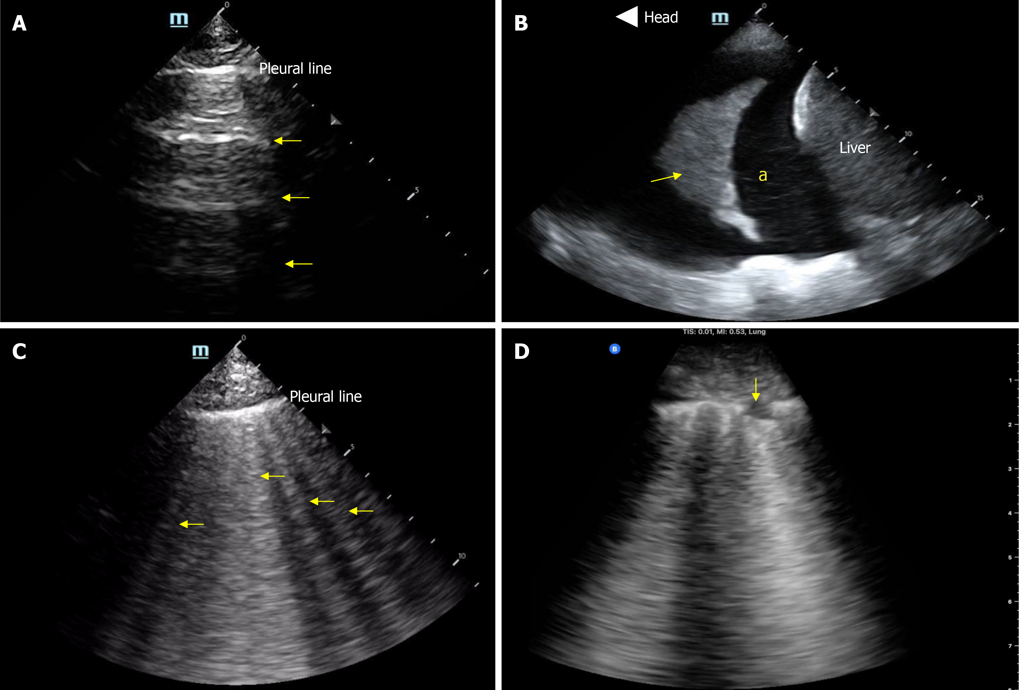
Figure 3 Basic lung ultrasound findings.
A: Normal lung with horizontal hyperechoic artifacts known as A-lines; B: Pleural effusion (asterisk) appearing as an anechoic area above the liver; the arrow points to atelectatic lung; C: Vertical hyperechoic artifacts called B-lines emerging from the pleural line, indicative of interstitial thickening typically due to fluid accumulation; D: Interstitial pneumonia, characterized by confluent B-lines and an irregular pleural line, with the arrow indicating a subpleural consolidation. Citation: Diniz H, Ferreira F, Koratala A. Point-of-care ultrasonography in nephrology: Growing applications, misconceptions and future outlook. World J Nephrol 2025; 25: 14: 105374. Copyright© The Author(s) 2025. Published by Baishideng Publishing Group Inc[29].
- Citation: Chandra NC, Bhattacharya D, Koratala A. Point-of-care ultrasound: Uniting cardiology and nephrology at the bedside. World J Cardiol 2025; 17(12): 112047
- URL: https://www.wjgnet.com/1949-8462/full/v17/i12/112047.htm
- DOI: https://dx.doi.org/10.4330/wjc.v17.i12.112047