Copyright
©The Author(s) 2023.
World J Cardiol. Dec 26, 2023; 15(12): 642-648
Published online Dec 26, 2023. doi: 10.4330/wjc.v15.i12.642
Published online Dec 26, 2023. doi: 10.4330/wjc.v15.i12.642
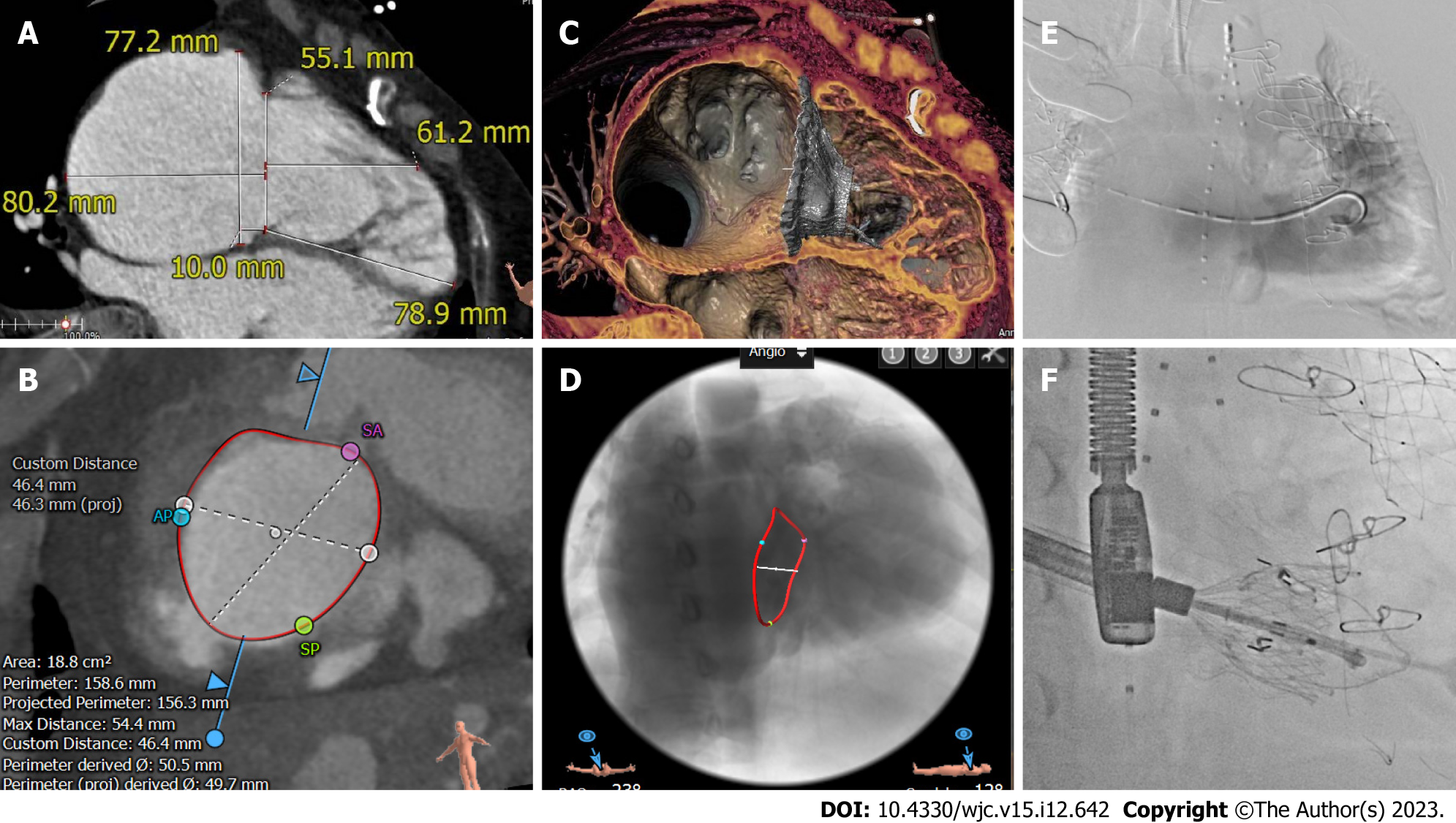
Figure 2 Preoperative computed tomography and intraoperative digital subtraction angiography images.
A and B: Computed tomography of the right ventricle revealed an enlarged right heart and tricuspid annulus; C and D: Simulating the morphology of the prosthetic tricuspid valve after placement; E: The guide wire passed through the tricuspid valve into the right ventricle and angiography indicated massive tricuspid regurgitation; F: Release of the artificial tricuspid valve.
- Citation: Cao JY, Ning XP, Zhou GW, Li BL, Qiao F, Han L, Xu ZY, Lu FL. Pulmonary and tricuspid regurgitation after Tetralogy of Fallot repair: A case report. World J Cardiol 2023; 15(12): 642-648
- URL: https://www.wjgnet.com/1949-8462/full/v15/i12/642.htm
- DOI: https://dx.doi.org/10.4330/wjc.v15.i12.642


