
















©The Author(s) 2020.
World J Cardiol. May 26, 2020; 12(5): 167-191
Published online May 26, 2020. doi: 10.4330/wjc.v12.i5.167
Published online May 26, 2020. doi: 10.4330/wjc.v12.i5.167
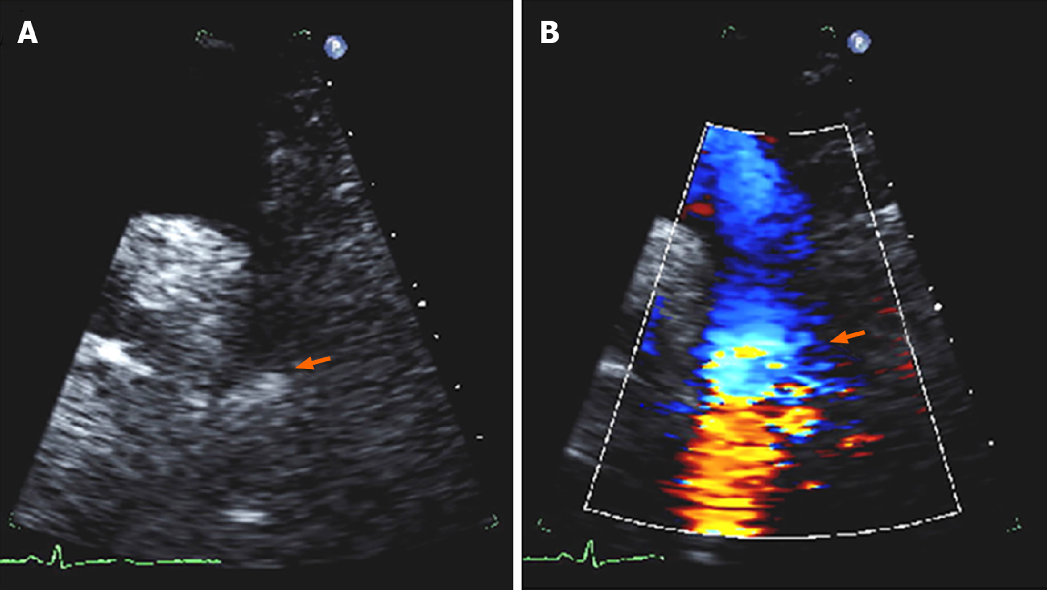
Figure 6 Echocardiographic findings in coarctation of aorta.
A: Two-dimensional suprasternal view on transthoracic echocardiogram demonstrating narrowing in the aortic lumen at the isthmus (arrow); B: Color Doppler imaging demonstrating turbulent flow at the site of the coarctation (arrow).
- Citation: Agasthi P, Pujari SH, Tseng A, Graziano JN, Marcotte F, Majdalany D, Mookadam F, Hagler DJ, Arsanjani R. Management of adults with coarctation of aorta. World J Cardiol 2020; 12(5): 167-191
- URL: https://www.wjgnet.com/1949-8462/full/v12/i5/167.htm
- DOI: https://dx.doi.org/10.4330/wjc.v12.i5.167