Copyright
©The Author(s) 2017.
World J Cardiol. Aug 26, 2017; 9(8): 673-684
Published online Aug 26, 2017. doi: 10.4330/wjc.v9.i8.673
Published online Aug 26, 2017. doi: 10.4330/wjc.v9.i8.673
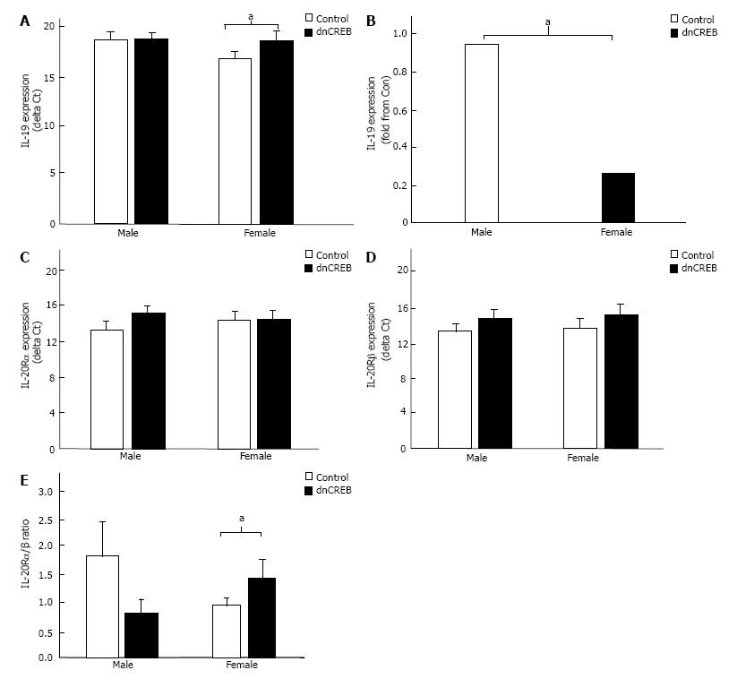
Figure 1 Expression of interleukin-19 and interleukin-20R in the dominant negative cyclic AMP response-element binding protein model of female-dominant heart failure.
A: Female male significantly downregulated IL-19 expression in the setting of dnCREB-mediated heart failure compared to male mice which maintained IL-19 expression; B: A significant difference between sexes; C: IL-20Rα and IL-20Rβ (D) expression were unchanged in either sex; D: However, the ratio between IL-20Rα/β was significantly upregulated in female dnCREB mice. Expression of IL-19 and IL-20R was assessed by qRT-PCR, and ΔCt calculated relative to 18S. The ratio of IL-20Rα/β was calculated as a fold change of α-β within each sex. Data are expressed as mean ± SEM. aP < 0.05, n = 5-9 mice per group. dnCREB: Do-minant negative cyclic AMP response-element binding protein.
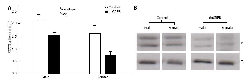
Figure 2 Activation of signal transducer and activator of transcription 3 in the dominant negative cyclic AMP response-element binding protein model of female-dominant heart failure.
A: Male and female dnCREB mice show attenuated STAT3 activation compared to controls; B: Representative immunoblotting images. Activation of STAT3 was assessed by immunoblotting of phospho STAT3/total STAT3. Data are expressed as mean ± SEM and analyzed by 2-way ANOVA. All four conditions were run on the same gel, and non-essential lanes were removed for generation of the representative images. aP < 0.05, n = 2-6 mice per group. a: Significant effect of genotype. dnCREB: Do-minant negative cyclic AMP response-element binding protein; STAT3: Signal transducer and activator of transcription 3.
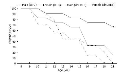
Figure 3 Kaplan-Meier survival curve of dominant negative cyclic AMP response-element binding protein and double transgenic male and female mice.
IL-19 knockout in the setting of dnCREB accelerates male mouse mortality, while not affecting female dnCREB survival. No mortality was observed in Control or IL-19 KO mice during this period. Log-rank analyses were performed to compare survival between groups. aP < 0.05 vs female DTG, female dnCREB, and male DTG; n = 7-10 mice per group. dnCREB: Do-minant negative cyclic AMP response-element binding protein; DTG: Double transgenic.
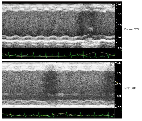
Figure 4 Representative images from M-mode echocardiographic analysis of cardiac function in male and female double transgenic mice.
Quantification of echocardiographic analyses are presented in Table 1. DTG: Double transgenic.
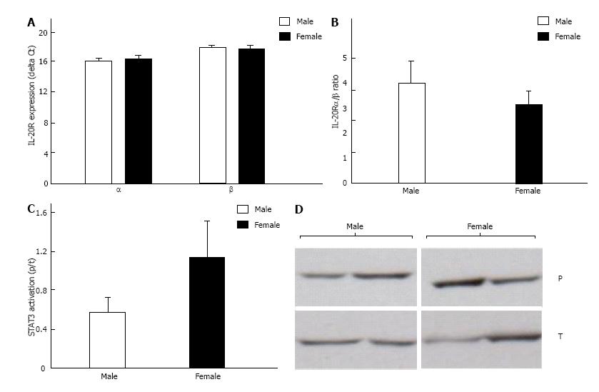
Figure 5 Expression of interleukin-20Rα/β and activation of signal transducer and activator of transcription 3 in male and female double transgenic mice.
A: IL-20Rα and IL-20Rβ expression were similar in male and female DTG mice; B: Similarly, the ratio of IL-20Rα/β did not differ between sexes; C: STAT3 activation did not differ between male and female DTG mice; D: Representative immunoblotting images. Expression of IL-20R was assessed by qRT-PCR and expressed and ΔCt calculated relative to 18S. The ratio of IL-20Rα/β was calculated as a fold change of α-β within each sex. Activation of STAT3 was assessed by immunoblotting of phospho STAT3/total STAT3. Both male and female mice were run on the same gel, and non-essential lanes were cropped for generation of the representative image. Data are expressed as mean ± SEM; n = 4-7 mice per group. STAT3: Signal transducer and activator of transcription 3; DTG: Double transgenic.
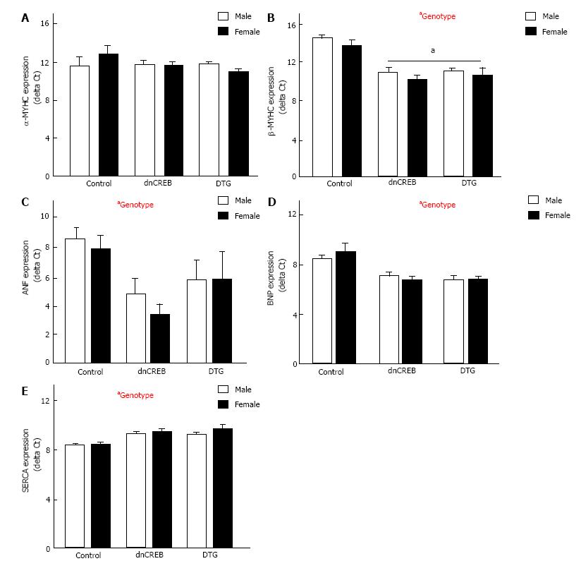
Figure 6 Activation of the fetal hypertrophic gene program in dominant negative cyclic AMP response-element binding protein and double transgenic mice.
A: Myosin heavy chain α (MYHCα) expression was unchanged by either sex or genotype; B: Myosin heavy chain β (MYHCβ); C: Atrial natriuretic factor (ANF); D: Brain natriuretic peptide (BNP) were all significantly upregulated in both male and female dnCREB and DTG mice; E: Sarcoplasmic reticulum Ca2+ ATPase (SERCA) expression was significantly downregulated in both sexes with disease. Expression of fetal hypertrophic genes was assessed by qRT-PCR, relative to the housekeeping gene 18S. Data are expressed as mean ± SEM and assessed by 2-way ANOVA. aP < 0.05 vs Control; n = 4-7 mice per group. a: Significant effect of genotype. dnCREB: Do-minant negative cyclic AMP response-element binding protein; DTG: Double transgenic.
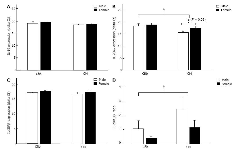
Figure 7 Interleukin-19 and interleukin-20R expression in male and female cardiac fibroblasts and myocytes.
A: Both male and female Cfib and CM express IL-19, with no differences between sexes or cell types; B: Both Cfib and CM express IL-20Rα, with higher expression in CM than Cfib, as evident by the lower delta Ct. Male CM tended to express higher IL-20Rα than female CM (P = 0.06); C: Both Cfib and CM express IL-20Rβ, with no differences between cell types or sex; D: The ratio of IL-20Rα/β was significantly higher in CM than Cfib. IL-19 and IL-20R expression were assessed by qRT-PCR and normalized to the housekeeping gene 18S. Data are expressed as mean ± SEM and assessed by 2-way ANOVA; n = 7-8 mice per group. Cfib: Cardiac fibroblasts; CM: Cardiac myocytes.
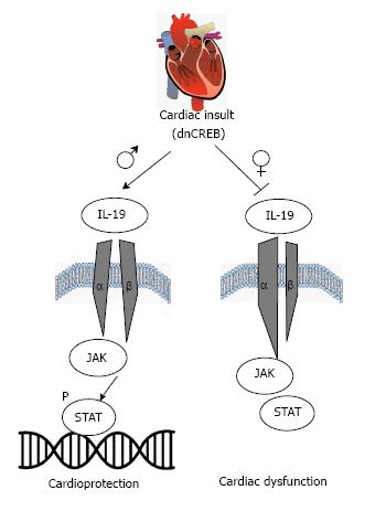
Figure 8 Working model of interleukin-19 in sex-specific heart failure.
Cardiac insult (inactivation of cyclic AMP response-element binding protein) results in maintained interleukin-19 (IL-19) expression in male mice, with maintained expression of IL-20 receptor subunits. Activation of downstream JAK-STAT signaling in male hearts is cardioprotective. However, cardiac insult in female mice results in attenuated IL-19 signaling, dysregulated expression of IL-20R subunits, and cardiac dysfunction. Ongoing investigations will delineate the downstream mediators implicated in IL-19 cardioprotection in a sex-specific manner.
- Citation: Bruns DR, Ghincea AR, Ghincea CV, Azuma YT, Watson PA, Autieri MV, Walker LA. Interleukin-19 is cardioprotective in dominant negative cyclic adenosine monophosphate response-element binding protein-mediated heart failure in a sex-specific manner. World J Cardiol 2017; 9(8): 673-684
- URL: https://www.wjgnet.com/1949-8462/full/v9/i8/673.htm
- DOI: https://dx.doi.org/10.4330/wjc.v9.i8.673


