Copyright
©The Author(s) 2016.
World J Biol Chem. Feb 26, 2016; 7(1): 168-177
Published online Feb 26, 2016. doi: 10.4331/wjbc.v7.i1.168
Published online Feb 26, 2016. doi: 10.4331/wjbc.v7.i1.168
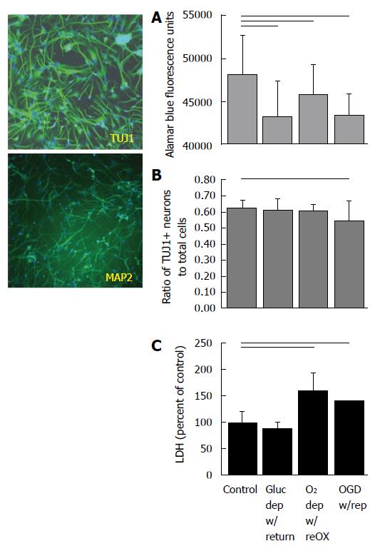
Figure 1 Cultures of human neuronal progenitor cells express TUJ1 and MAP2ab at 14 d in vitro (left) and die within 24 h of re-oxygenation (right).
Cultures were deprived of oxygen and/or glucose for 48 h and resources were replaced for 24 h before assaying for viability (A), proportion NPC to total cells (B) or cell death (C). Horizontal lines indicate P < 0.05; ANOVA followed by a Holm-Sidak test (A, B) or Kruskal-Wallis ANOVA on ranks followed by Dunn’s multiple comparisons (C). Mean ± SD. Only differences relative to control are shown. NPC: Neuronal progenitor cell.
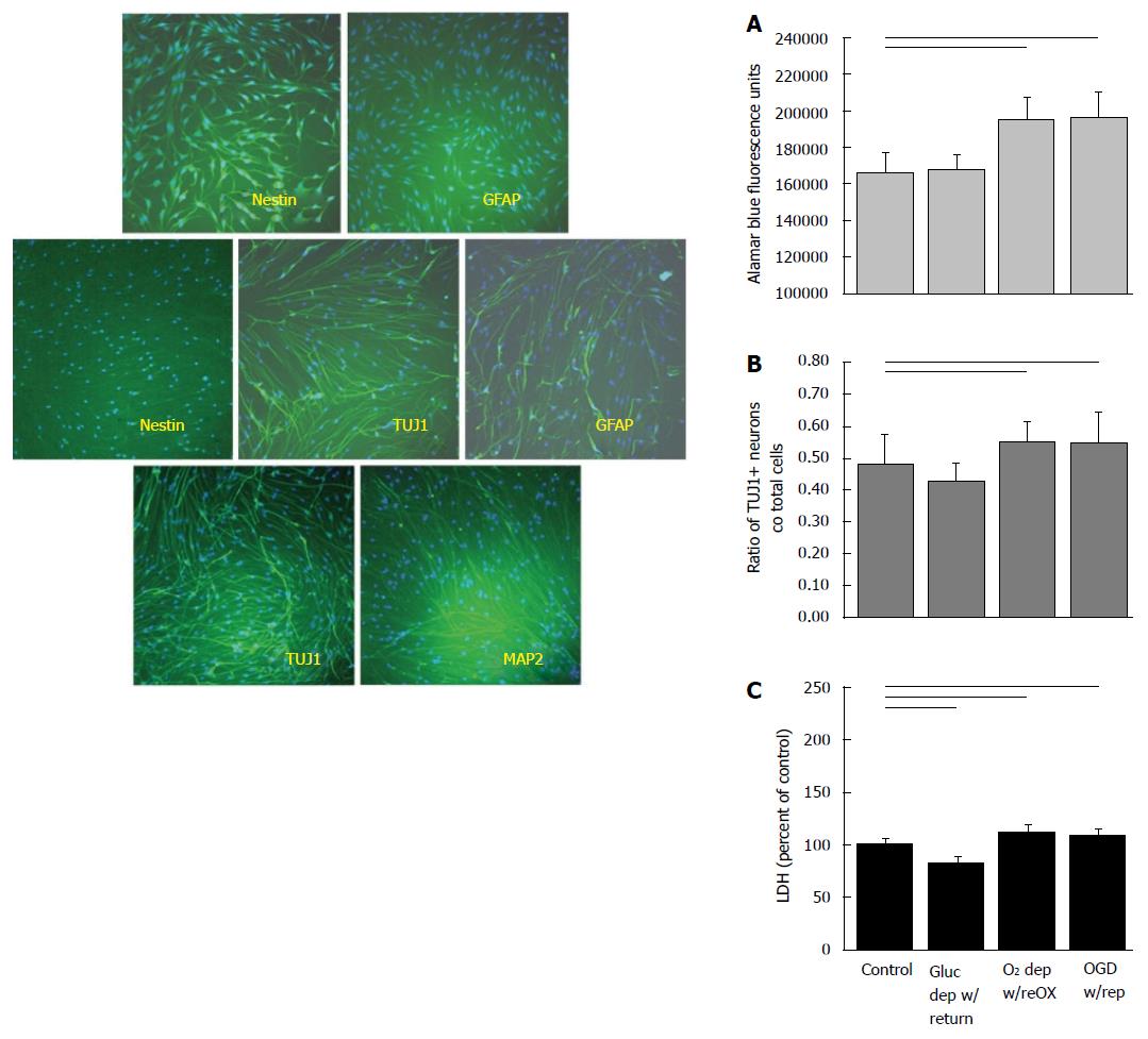
Figure 2 Cultures of arctic ground squirrel neuronal progenitor cells do not begin to express MAP2ab, characteristic of mature neurons, until 21 d in vitro.
Cultures of AGS NPC express nestin, a marker of NSC, but not GFAP, a marker for astrocytes at 7 d in vitro (DIV) (top left). By 14 DIV cultures express TUJ1 a marker of neuronal progenitor cells that are committed to a neuronal fate, and GFAP a marker of progenitor cells commitment to an astrocytic fate. Nestin is no longer expressed at 14 DIV (middle left). At 21 DIV cultures express TUJ1 and begin to express MAP2ab (bottom left). AGS NPCs at 14 DIV increase in viability and appear to proliferate when deprived of oxygen or oxygen and glucose (right). When AGS NPC were deprived of oxygen or oxygen and glucose for 48 h followed by 24 h of resource replacement, cultures showed an increase in viability (A) and an increase in the proportion of neuronal progenitors (B) despite signs of cell death (C). Horizontal lines P < 0.05; ANOVA followed by a Holm-Sidak test. Mean ± SD. Only differences relative to control are shown. AGS: Arctic ground squirrel; NPC: Neuronal progenitor cell; OGD: Oxygen glucose deprivation.
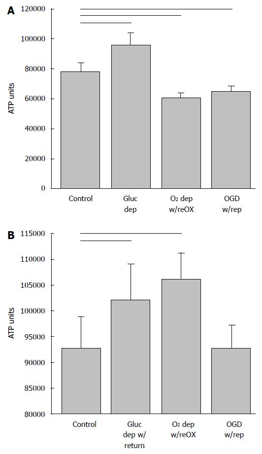
Figure 3 ATP (shown as relative luminescence units) declines in arctic ground squirrel neuronal progenitor cells during oxygen and glucose deprivation.
NPCs at 14 d in vitro deprived for 48 h followed by 24 h resource replacement lost ATP when deprived of oxygen or oxygen and glucose (A). ATP recovered within 24 h of reoxygenation or reperfusion (modeled by return of oxygen and glucose) (B). Horizontal lines P < 0.05; ANOVA followed by a Holm-Sidak test. Mean + SD. Only differences relative to control are shown. OGD: Oxygen and glucose deprivation; NPCs: Neuronal progenitor cells.
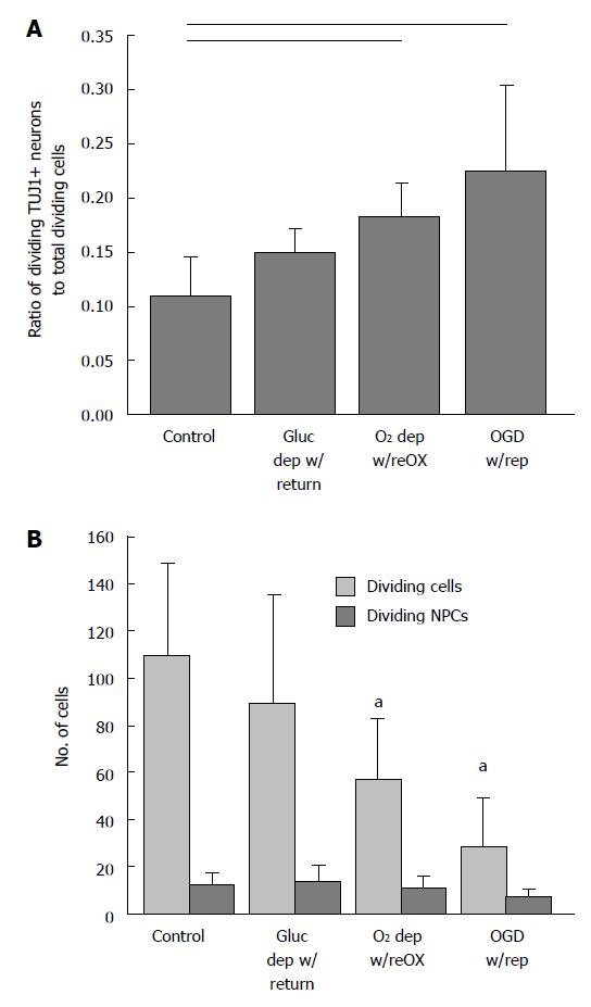
Figure 4 Arctic ground squirrel neuronal progenitor cells proliferate in response to injury faster than other cell lineages.
AGS NPCs at 14 d in vitro deprived of oxygen or oxygen and glucose for 48 h followed by 24 h of reoxygenation or reperfusion (modeled by return of oxygen and glucose). The proportion of dividing NPCs (TUJ1+ and Ki67+/Ki67+) (A) increased because the number of dividing NPCs (TUJ1+ and Ki67+) did not change while the number of dividing cells (Ki67+) decreased (B). Horizontal lines P < 0.05; aP < 0.05 compared to control; ANOVA followed by a Holm-Sidak test. Mean ± SD. Only differences relative to control are shown. OGD: Oxygen glucose deprivation; NPCs: Neuronal progenitor cells; AGS: Arctic ground squirrel.
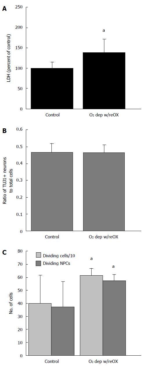
Figure 5 Arctic ground squirrel neuronal progenitor cell cultured for 21 d in vitro are more vulnerable to oxygen deprivation than cells cultured for 14 d in vitro.
Longer time in culture lowers resistance to oxygen deprivation /reoxygenation indicated by an increase in LDH (A) not seen after 14 d in vitro (DIV). However, after 24 h of O2 deprivation, followed by 24 h of reoxygenation the proportion of neuronal to nonneuronal progenitors did not change (B) while proliferation of NPCs increased (C). Data are from AGS NPCs cultured for 21 DIV that were deprived of oxygen followed by 24 h of reoxygenation. aP < 0.05 compared to control, t-test. LDH: Lactate dehydrogenase; NPCs: Neuronal progenitor cells; AGS: Arctic ground squirrel.
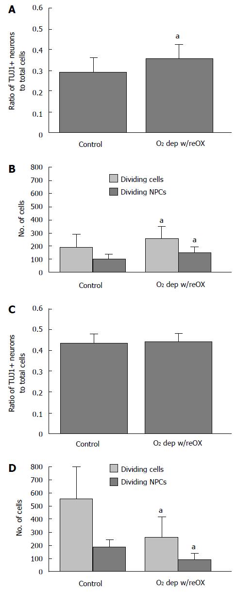
Figure 6 By 48 and 72 h after re-oxygenation the ratio of neuronal progenitor cells to total cells increases slightly or remains the same.
AGS NPCs cultured for 21 d in vitro were deprived of oxygen followed by 48 h (A, B) or 72 h (C, D) of re-oxygenation. After 48 h of reoxygenation, the proportion of NPCs increased relative to total cells because the number of dividing NPCs increased (B). After 72 h the proportion of NPCs was sustained (C) because division of NPCs declined less than other cell lineages (D). aP < 0.05 compared to control, t-test. Mean ± SD. AGS: Arctic ground squirrel; NPCs: Neuronal progenitor cells.
Figure 7 Arctic ground squirrel neuronal progenitor cells are vulnerable to oxidative stress.
Cells, 14 d in vitro, were exposed to 50 μmol/L H2O2. After 2 to 8 h of exposure cellular respiration, a measure of cellular viability, was measured using alamarBlue®. The viability of human cultures (A) decreased after 6 h exposure while the viability of AGS NPCs (B) decreased after 2 h of exposure to H2O2. AGS: Arctic ground squirrel; NPCs: Neuronal progenitor cells.
- Citation: Drew KL, Wells M, McGee R, Ross AP, Kelleher-Andersson J. Arctic ground squirrel neuronal progenitor cells resist oxygen and glucose deprivation-induced death. World J Biol Chem 2016; 7(1): 168-177
- URL: https://www.wjgnet.com/1949-8454/full/v7/i1/168.htm
- DOI: https://dx.doi.org/10.4331/wjbc.v7.i1.168


