Copyright
©The Author(s) 2017.
World J Gastrointest Oncol. Sep 15, 2017; 9(9): 397-401
Published online Sep 15, 2017. doi: 10.4251/wjgo.v9.i9.397
Published online Sep 15, 2017. doi: 10.4251/wjgo.v9.i9.397
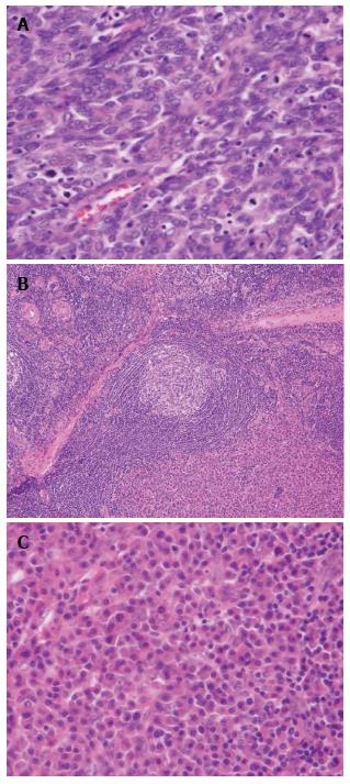
Figure 5 Histopathological results of the resected specimen.
A: The histopathological diagnosis of the resected esophagus is moderately differentiated squamous cell carcinoma (HE × 200); B: Histological examination of the right-sided neck lymph nodes reveals onionskin arrangement of small lymphocytes (HE × 20); C: Interfollicular diffuse proliferation of plasma cells (HE × 200).
- Citation: Yamabuki T, Ohara M, Kato M, Kimura N, Shirosaki T, Okamura K, Fujiwara A, Takahashi R, Komuro K, Iwashiro N, Hirano S. Cervical Castleman’s disease mimicking lymph node metastasis of esophageal carcinoma. World J Gastrointest Oncol 2017; 9(9): 397-401
- URL: https://www.wjgnet.com/1948-5204/full/v9/i9/397.htm
- DOI: https://dx.doi.org/10.4251/wjgo.v9.i9.397


