Copyright
©2013 Baishideng Publishing Group Co.
World J Gastrointest Oncol. Jul 15, 2013; 5(7): 115-126
Published online Jul 15, 2013. doi: 10.4251/wjgo.v5.i7.115
Published online Jul 15, 2013. doi: 10.4251/wjgo.v5.i7.115
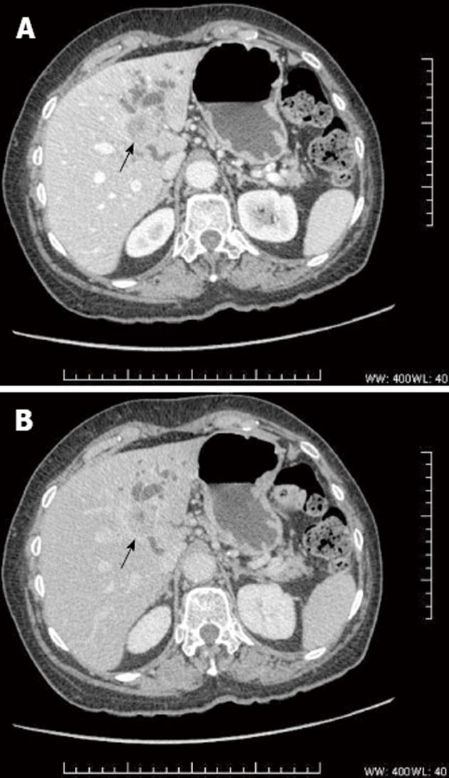
Figure 1 Mass forming cholangiocarcinoma.
Axial computed tomography scan in the portal phase (A) and in delayed phase (B) shows a heterogeneous hypovascular mass (arrow) with rim-like peripheral enhancement. The lesion is associated with ductal infiltration and biliary dilation.
- Citation: Valls C, Ruiz S, Martinez L, Leiva D. Radiological diagnosis and staging of hilar cholangiocarcinoma. World J Gastrointest Oncol 2013; 5(7): 115-126
- URL: https://www.wjgnet.com/1948-5204/full/v5/i7/115.htm
- DOI: https://dx.doi.org/10.4251/wjgo.v5.i7.115


