Copyright
©2013 Baishideng Publishing Group Co.
World J Gastrointest Oncol. Dec 15, 2013; 5(12): 222-229
Published online Dec 15, 2013. doi: 10.4251/wjgo.v5.i12.222
Published online Dec 15, 2013. doi: 10.4251/wjgo.v5.i12.222
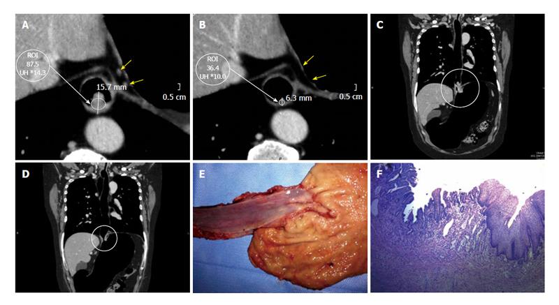
Figure 6 Advanced adenocarcinoma of the gastroesophageal junction.
Dworak grade 2, large areas of fibrosis or necrosis with active cells. A: Axial pre-therapy image. Both wall thickness and density are measured. Yellow arrows indicate two small lymph nodes; B: Axial post-therapy image reveals a clear decrease of tumor size and in density. Lymph nodes are not visible; C: Coronal multiplanar reconstructions (MPR) pre-therapy reconstruction. The circle shows the long axis of the tumor; D: Coronal MPR after therapy reconstruction. Note marked decrease in tumor size; E: Surgical specimen of total esophagectomy and upper polar gastrectomy correlating well with the lesion demonstrated with pneumo-computed tomography. Open piece, is recognized at the level of the gastro-esophageal junction an indurated area with diminished mucosal folds and elevated edges with whitish nodule and firm consistency; F: Submucosal layer with nodular accumulations of atypical epithelial cells scant cytoplasm that are arranged in small tubular structures, cords or dispersed. Nodules are found at the distal esophagus and cardia underlying mucosa. Linfovasculares tumor emboli are observed. In small area is observed submucosal fibrosis, chronic inflammation and congestion. At the level of the gastroesophageal junction is observed extensive intestinal metaplasia to intramucosal carcinoma focus. Preserved oxyntic gastric mucosa.
- Citation: Ulla M, Gentile E, Yeyati EL, Diez ML, Cavadas D, Garcia-Monaco RD, Ros PR. Pneumo-CT assessing response to neoadjuvant therapy in esophageal cancer: Imaging-pathological correlation. World J Gastrointest Oncol 2013; 5(12): 222-229
- URL: https://www.wjgnet.com/1948-5204/full/v5/i12/222.htm
- DOI: https://dx.doi.org/10.4251/wjgo.v5.i12.222


