Copyright
©2013 Baishideng Publishing Group Co.
World J Gastrointest Oncol. Dec 15, 2013; 5(12): 222-229
Published online Dec 15, 2013. doi: 10.4251/wjgo.v5.i12.222
Published online Dec 15, 2013. doi: 10.4251/wjgo.v5.i12.222
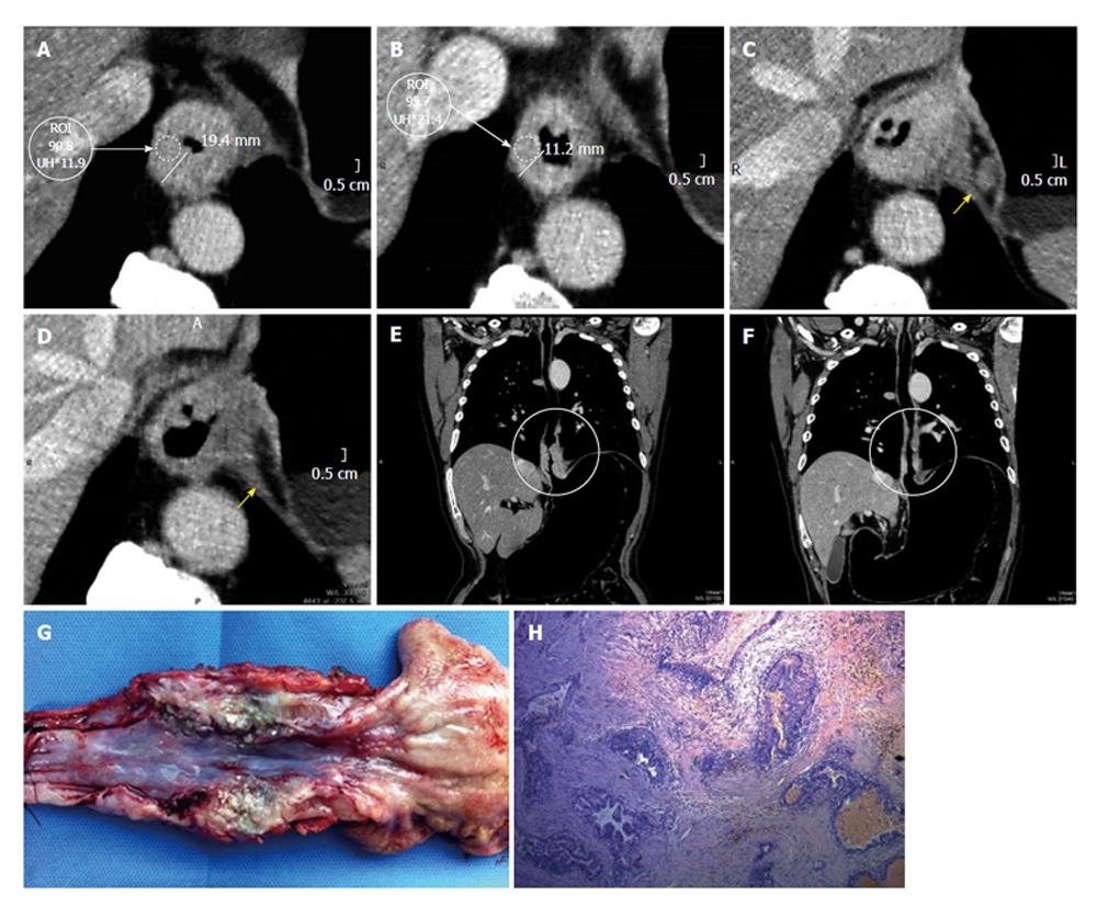
Figure 5 Advanced adenocarcinoma.
Dworak grade I, prevalence of active cells. A: Axial pre neoadjuvant therapy image. Both wall thickness and density are measured; B: Axial post-neoadjuvant therapy image reveals persistence of the wall thickening and almost no variation in density; C: Axial pre-neoadjuvant therapy image, the arrow is pointing to a lymphadenopathy; D: Axial post-neoadjuvancy image reveals resolution of the adenopathy; E: Coronal multiplanar reconstructions (MPR) pre-neoadjuvancy reconstruction. The circle shows the long axis compromise of the tumor; F: Coronal MPR post-therapy reconstruction. The circle shows the long axis of the tumor and its precise location; G: Surgical specimen of total esophagectomy and upper polar gastrectomy. Open piece shows an important thickening of the lower esophagus, at squamo-columnar junction recognizes a polypoid lesion and poorly defined borders. The remaining gastric mucosa presents edematous folds; H: Section shows at gastroesophageal junction one degrade injuries ranging from low-grade dysplasia to invasive adenocarcinoma. The primary lesion is below the squamocolumnar junction. Generally infiltrates shaped surface (submucosa) with occasional foci in muscle layer and extends in the form of multiple separate foci below esophageal epithelium. Proximal margin: adenocarcinoma foci observed at adventitia and submucosa layers. Tumor is seen infiltrating striated muscle tissue.
- Citation: Ulla M, Gentile E, Yeyati EL, Diez ML, Cavadas D, Garcia-Monaco RD, Ros PR. Pneumo-CT assessing response to neoadjuvant therapy in esophageal cancer: Imaging-pathological correlation. World J Gastrointest Oncol 2013; 5(12): 222-229
- URL: https://www.wjgnet.com/1948-5204/full/v5/i12/222.htm
- DOI: https://dx.doi.org/10.4251/wjgo.v5.i12.222


