Copyright
©2010 Baishideng.
World J Gastrointest Oncol. May 15, 2010; 2(5): 247-250
Published online May 15, 2010. doi: 10.4251/wjgo.v2.i5.247
Published online May 15, 2010. doi: 10.4251/wjgo.v2.i5.247
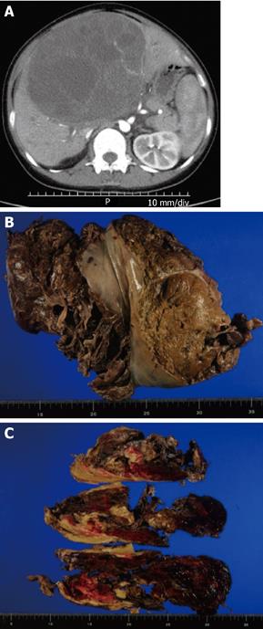
Figure 1 Computed tomography (CT) image and resected specimen.
A: Abdominal CT image before surgery; B: Resected specimen. The liver was ruptured; C: Cutting of the surface revealed a soft white mass with multilocular appearance, fibrous pseudocapsules, and massive hemorrhage.
- Citation: Kinjo S, Sakurai S, Hirato J, Sunose Y. Embryonal sarcoma of the liver with chondroid differentiation. World J Gastrointest Oncol 2010; 2(5): 247-250
- URL: https://www.wjgnet.com/1948-5204/full/v2/i5/247.htm
- DOI: https://dx.doi.org/10.4251/wjgo.v2.i5.247


