
















©2010 Baishideng.
World J Gastrointest Oncol. Mar 15, 2010; 2(3): 165-168
Published online Mar 15, 2010. doi: 10.4251/wjgo.v2.i3.165
Published online Mar 15, 2010. doi: 10.4251/wjgo.v2.i3.165
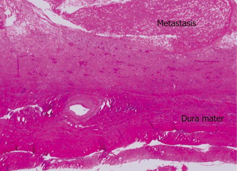
Figure 5 Dissection showed an 8 cm × 7 cm × 3 cm metastatic lesion had formed in the left occipitotemporal part of the cranial bone.
The lesion was osteolytic and showed invasion into the dura mater. Neither the subdural cavity nor the brain showed involvement from the metastatic tumor.
- Citation: Goto T, Dohmen T, Miura K, Ohshima S, Yoneyama K, Shibuya T, Kataoka E, Segawa D, Sato W, Anezaki Y, Ishii H, Kon D, Yamada I, Kamada K, Ohnishi H. Skull metastasis from hepatocellular carcinoma with chronic hepatitis B. World J Gastrointest Oncol 2010; 2(3): 165-168
- URL: https://www.wjgnet.com/1948-5204/full/v2/i3/165.htm
- DOI: https://dx.doi.org/10.4251/wjgo.v2.i3.165