Copyright
©2010 Baishideng.
World J Gastrointest Oncol. Feb 15, 2010; 2(2): 85-97
Published online Feb 15, 2010. doi: 10.4251/wjgo.v2.i2.85
Published online Feb 15, 2010. doi: 10.4251/wjgo.v2.i2.85
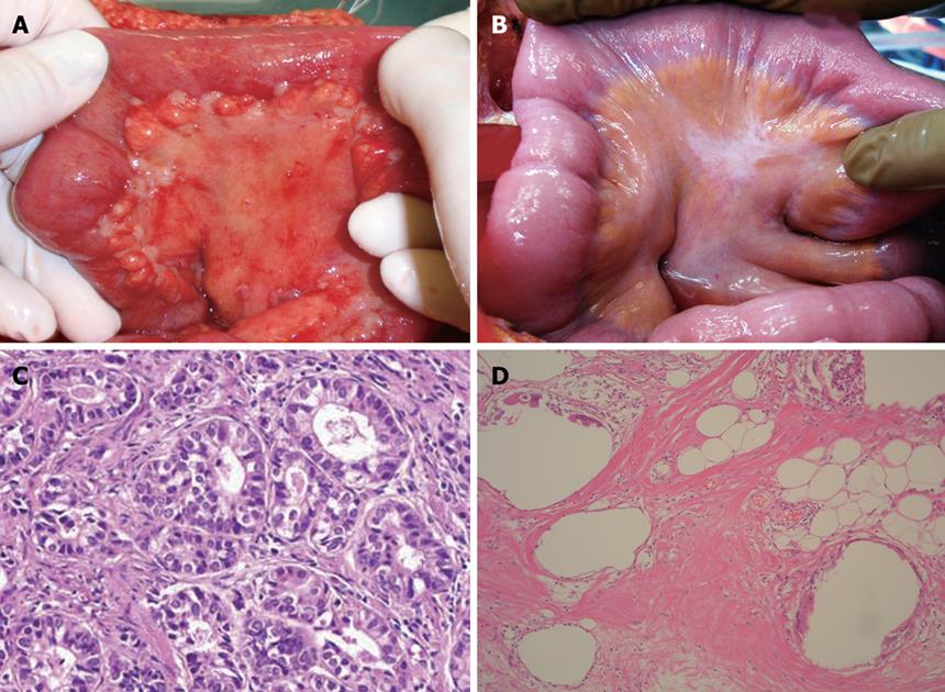
Figure 4 A 48-year old male patient with PC from gastric cancer treated with NIPS.
A: Macroscopic finding of PC on bowel mesentery; B: After 2 courses of NIPS, PC nodules shows fibrotic changes; C: Histologic findings of PC nodule obtained at the first operation of Figure 4A; D: Complete degeneration of cancer cells in PC nodule obtained at second look operation after NIPS.
- Citation: Yonemura Y, Elnemr A, Endou Y, Hirano M, Mizumoto A, Takao N, Ichinose M, Miura M, Li Y. Multidisciplinary therapy for treatment of patients with peritoneal carcinomatosis from gastric cancer. World J Gastrointest Oncol 2010; 2(2): 85-97
- URL: https://www.wjgnet.com/1948-5204/full/v2/i2/85.htm
- DOI: https://dx.doi.org/10.4251/wjgo.v2.i2.85


