
















©The Author(s) 2025.
World J Gastrointest Oncol. Sep 15, 2025; 17(9): 105937
Published online Sep 15, 2025. doi: 10.4251/wjgo.v17.i9.105937
Published online Sep 15, 2025. doi: 10.4251/wjgo.v17.i9.105937
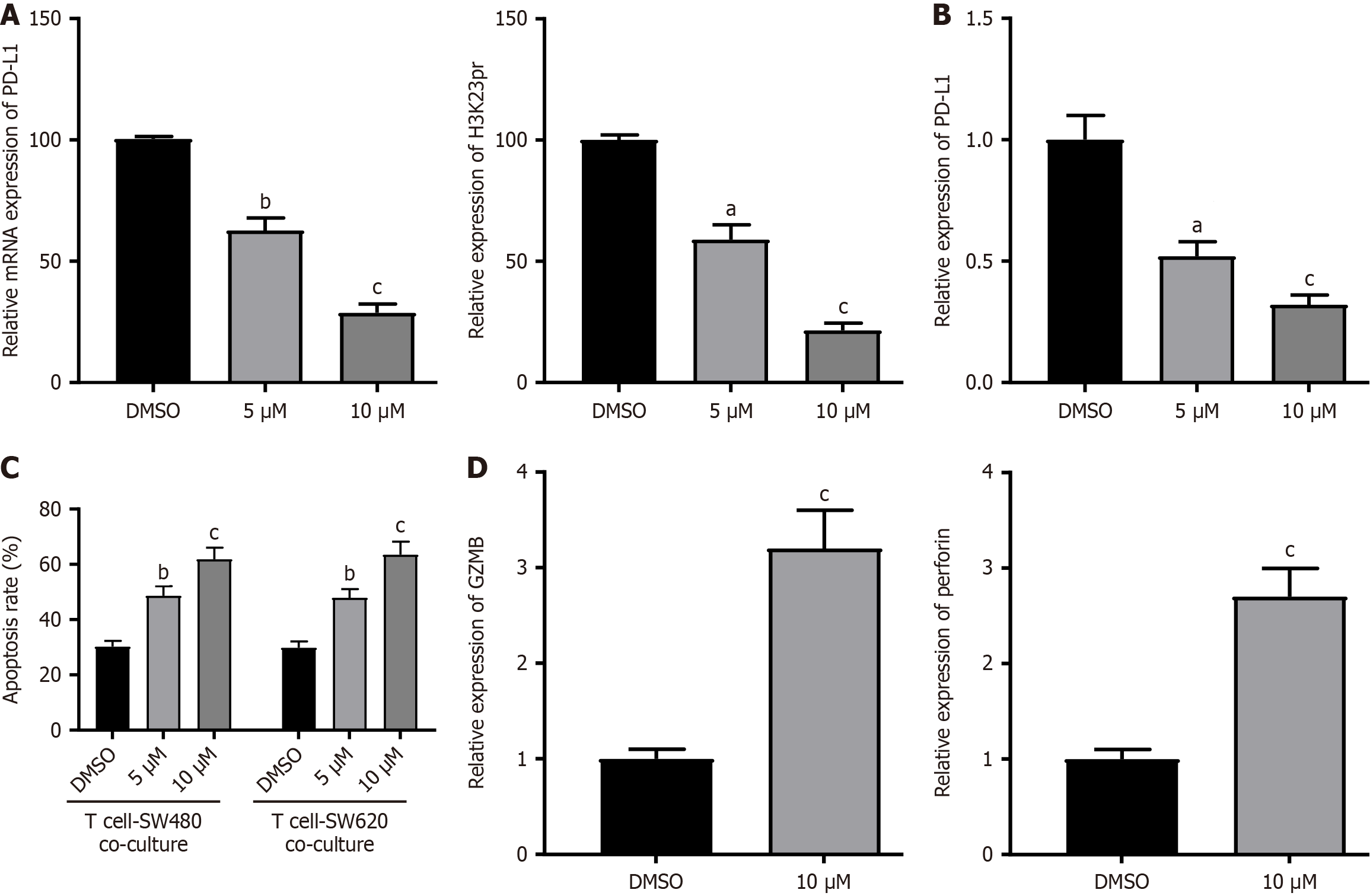
Figure 5 Pharmacological inhibition of KAT6A recapitulates genetic knockdown phenotypes.
A: Dose-dependent suppression of H3K23pr and PD-L1 by WM-3835. Western blot (left) and densitometric quantification (right) showed H3K23pr and PD-L1 protein levels in SW480/SW620 cells treated with WM-3835 (0, 5, or 10 μM) for 48 hours; B: PD-L1 transcriptional downregulation as revealed by quantitative PCR analysis of PD-L1 mRNA levels; C: Enhanced T cell-mediated apoptosis as shown by flow cytometry quantification of apoptotic colorectal cancer cells co-cultured with activated T cells; D: Activation of T cell cytotoxic markers demonstrated by Western blot analysis of GZMB and perforin in T cells. Data represent the mean ± SEM (n = 3). aP < 0.05, bP < 0.01, cP < 0.001 vs DMSO (one-way ANOVA with Dunnett’s test). siKAT6A data from Figures 1 and 2 are shown as positive controls.
- Citation: Zhou ZD, Zhao JP, Zheng SC, Wang TT. Correlation between KAT6A and PD-L1 expression and role of KAT6A in colorectal cancer. World J Gastrointest Oncol 2025; 17(9): 105937
- URL: https://www.wjgnet.com/1948-5204/full/v17/i9/105937.htm
- DOI: https://dx.doi.org/10.4251/wjgo.v17.i9.105937