Copyright
©The Author(s) 2025.
World J Gastrointest Oncol. Aug 15, 2025; 17(8): 107453
Published online Aug 15, 2025. doi: 10.4251/wjgo.v17.i8.107453
Published online Aug 15, 2025. doi: 10.4251/wjgo.v17.i8.107453
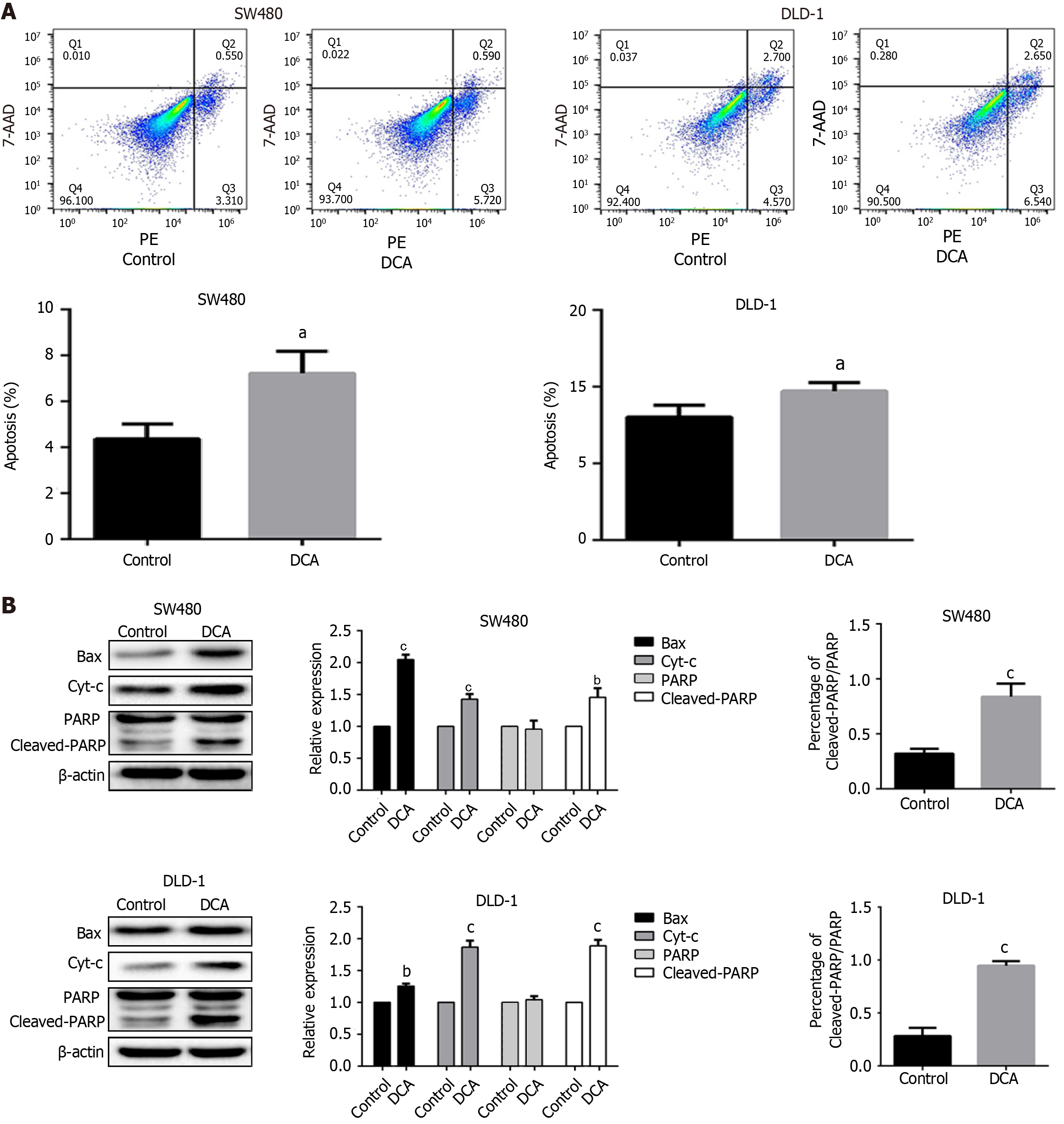
Figure 1 Deoxycholic acid promotes apoptosis in SW480 and DLD-1 cells.
A: Flow cytometry results and statistical plots for SW480 and DLD-1 cells; B: Western blot results and statistical plots of apoptosis-related proteins in SW480 and DLD-1 cells, and a histogram of the Cleaved-PARP/PARP ratio analysis. Control represents DMSO treatment, and the experimental group was treated with 100 μM deoxycholic acid for 24 hours. aP < 0.05, bP < 0.01, cP < 0.001. PE: Phycoerythrin; DCA: Deoxycholic acid.
- Citation: Chen JY, Wen JY, Lin JL, Li Y, Wu YZ, Lou LQ, Lou YL, Zuo ZG, Li X. Deoxycholic acid induces reactive oxygen species accumulation and promotes colorectal cancer cell apoptosis through the CaMKII-Ca2+ pathway. World J Gastrointest Oncol 2025; 17(8): 107453
- URL: https://www.wjgnet.com/1948-5204/full/v17/i8/107453.htm
- DOI: https://dx.doi.org/10.4251/wjgo.v17.i8.107453


