Copyright
©The Author(s) 2025.
World J Gastrointest Oncol. Jun 15, 2025; 17(6): 107272
Published online Jun 15, 2025. doi: 10.4251/wjgo.v17.i6.107272
Published online Jun 15, 2025. doi: 10.4251/wjgo.v17.i6.107272
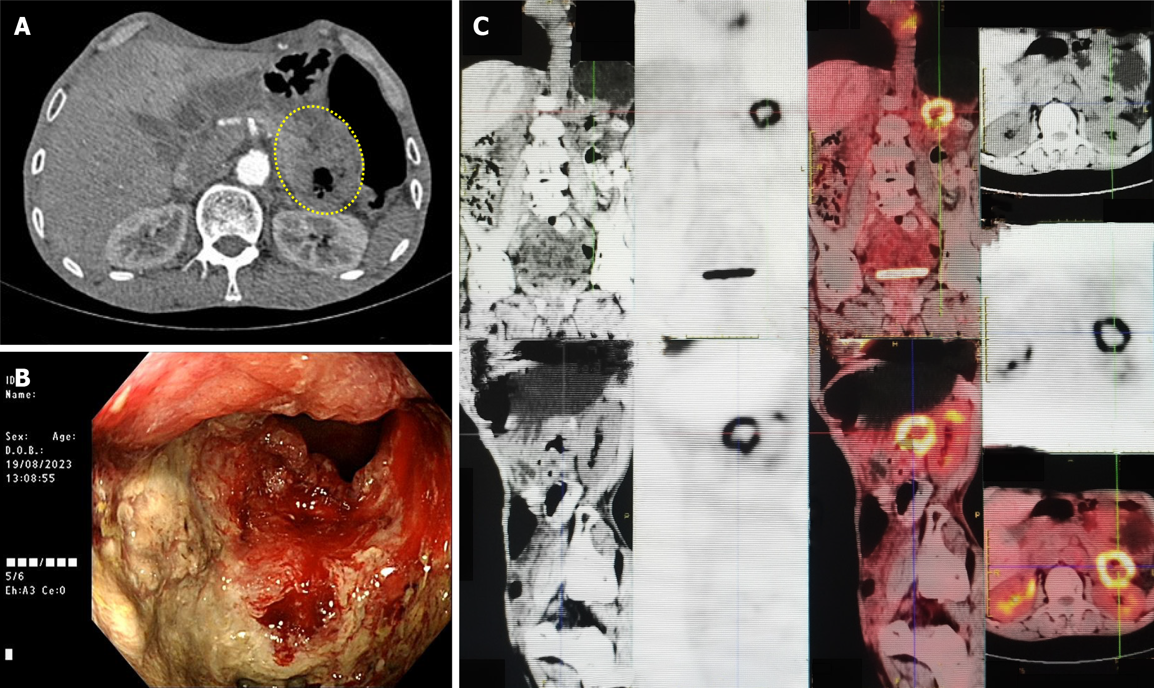
Figure 1 Imaging findings.
A: Computed tomography image showing circumferential wall thickening of the descending colon with luminal narrowing (yellow circle); B: Colonoscopic image of a circumferential ulcerative mass in the descending colon; C: Positron emission tomography-computed tomography image demonstrating hypermetabolic activity in the descending colon.
- Citation: Hu RR, Liu M, Li HY. Primary squamous cell carcinoma of the descending colon with pancreatic metastasis: A case report. World J Gastrointest Oncol 2025; 17(6): 107272
- URL: https://www.wjgnet.com/1948-5204/full/v17/i6/107272.htm
- DOI: https://dx.doi.org/10.4251/wjgo.v17.i6.107272


