Copyright
©The Author(s) 2025.
World J Gastrointest Oncol. May 15, 2025; 17(5): 102417
Published online May 15, 2025. doi: 10.4251/wjgo.v17.i5.102417
Published online May 15, 2025. doi: 10.4251/wjgo.v17.i5.102417
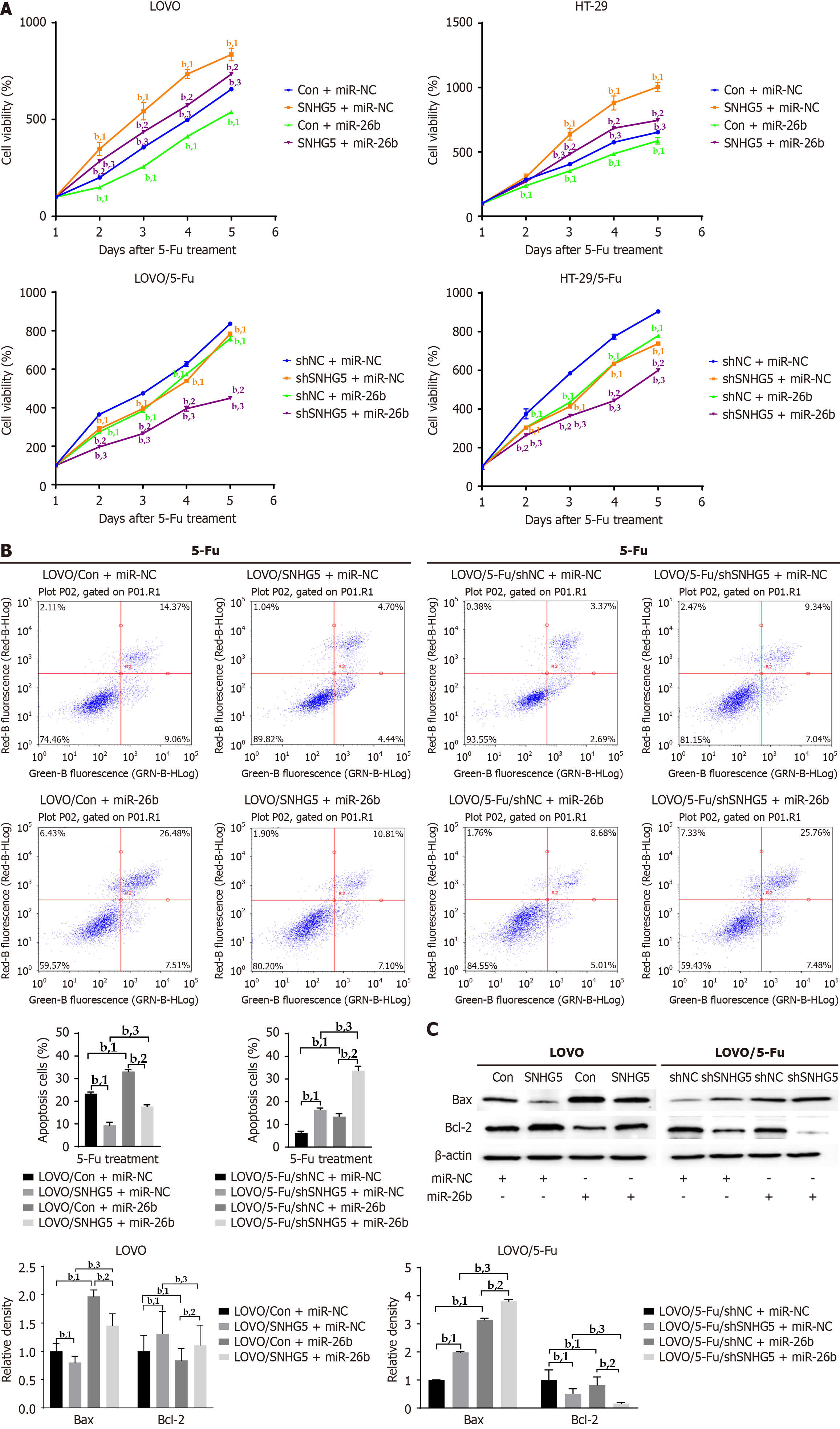
Figure 6 SNHG5 promotes colorectal cell resistance to 5-fluorouracil by sponging miR-26b.
The stable cells were transfected with miR-26b or miR-control (miR-NC), after 24 hours, and then treated with 5-fluorouracil. A: Cell viability was analyzed using cell counting kit 8 assays; B: The apoptosis rate of stable cells was analyzed using annexin-V fluorescein 5-isothiocyanate/propidium iodide staining assays (left) and quantified (right); C: Western blot analysis was performed to assess the expression of markers of apoptosis (Bax and Bcl-2), with β-actin serving as an internal control (left), and a densitometric analysis of Western blot signals is shown (right). bP < 0.01. 1P compared with the control + miR-NC group or the negative control-short hairpin RNA (shNC) + miR-NC group. 2P compared with the control + miR-26b group or the shNC + miR-26b group. 3P compared with the SNHG5 + miR-NC group or the SNHG5-short hairpin RNA + miR-NC group. miR-NC: MiR-control; Con: Control; shSNHG5: SNHG5-short hairpin RNA; shNC: Negative control-short hairpin RNA; 5-Fu: 5-fluorouracil.
- Citation: Wang B, Zhou Q, Cheng CE, Gu YJ, Jiang TW, Qiu JM, Wei GN, Feng YD, Ren LH, Shi RH. Long noncoding RNA SNHG5 promotes 5-fluorouracil resistance in colorectal cancer by regulating miR-26b/p-glycoprotein axis. World J Gastrointest Oncol 2025; 17(5): 102417
- URL: https://www.wjgnet.com/1948-5204/full/v17/i5/102417.htm
- DOI: https://dx.doi.org/10.4251/wjgo.v17.i5.102417


