
















©The Author(s) 2025.
World J Gastrointest Oncol. Oct 15, 2025; 17(10): 110570
Published online Oct 15, 2025. doi: 10.4251/wjgo.v17.i10.110570
Published online Oct 15, 2025. doi: 10.4251/wjgo.v17.i10.110570
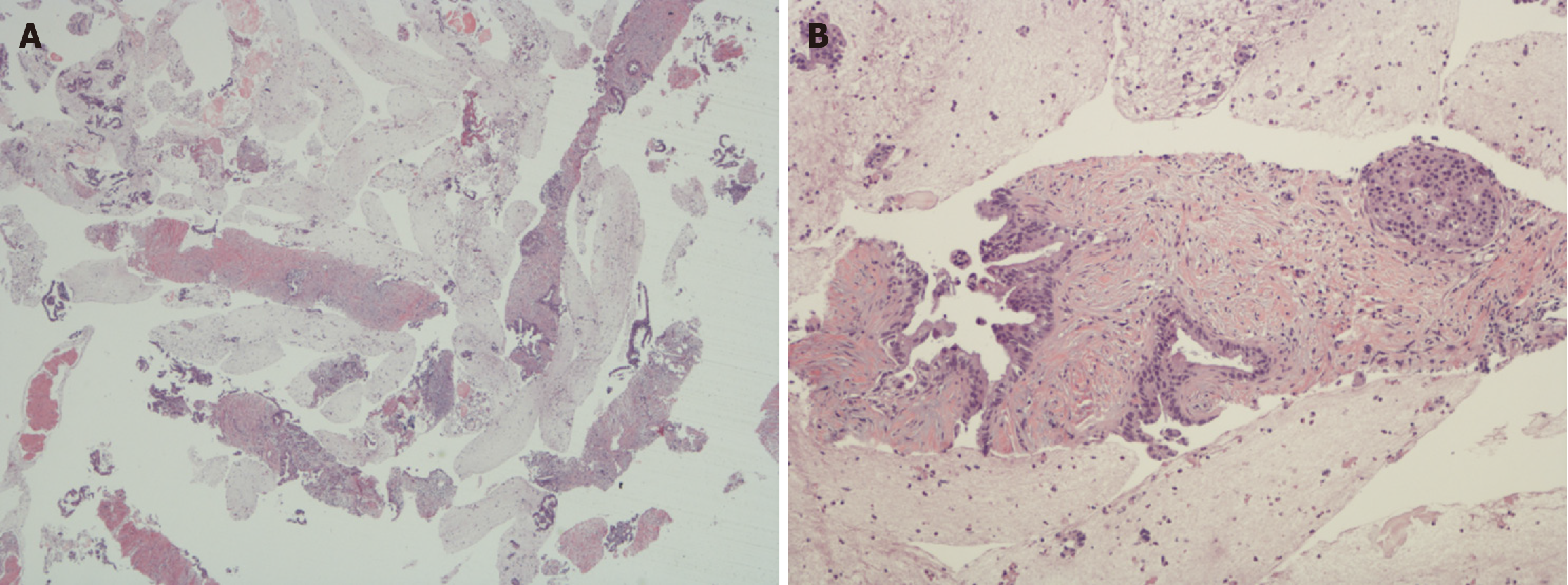
Figure 1 Photomicrographs of well-differentiated pancreatic ductal adenocarcinoma.
A: Low power view of fine needle biopsy that was obtained using endoscopic ultrasound. The glass slide showed > 2000 pancreatic ductal adenocarcinoma tumor cells which is very helpful for ancillary studies (original magnification × 20); B: Higher power view of one of the cores from Figure 1A showing a well-differentiated pancreatic ductal adenocarcinoma (the well-differentiated glands can be seen on the left and an islet of Langerhans on the right) (original magnification × 100).
- Citation: Moyana TN. Contrast-enhanced ultrasound as a non-invasive diagnostic modality for pancreatic ductal adenocarcinoma: The question of Ki67 for study validation. World J Gastrointest Oncol 2025; 17(10): 110570
- URL: https://www.wjgnet.com/1948-5204/full/v17/i10/110570.htm
- DOI: https://dx.doi.org/10.4251/wjgo.v17.i10.110570