Copyright
©The Author(s) 2024.
World J Gastrointest Oncol. Apr 15, 2024; 16(4): 1660-1667
Published online Apr 15, 2024. doi: 10.4251/wjgo.v16.i4.1660
Published online Apr 15, 2024. doi: 10.4251/wjgo.v16.i4.1660
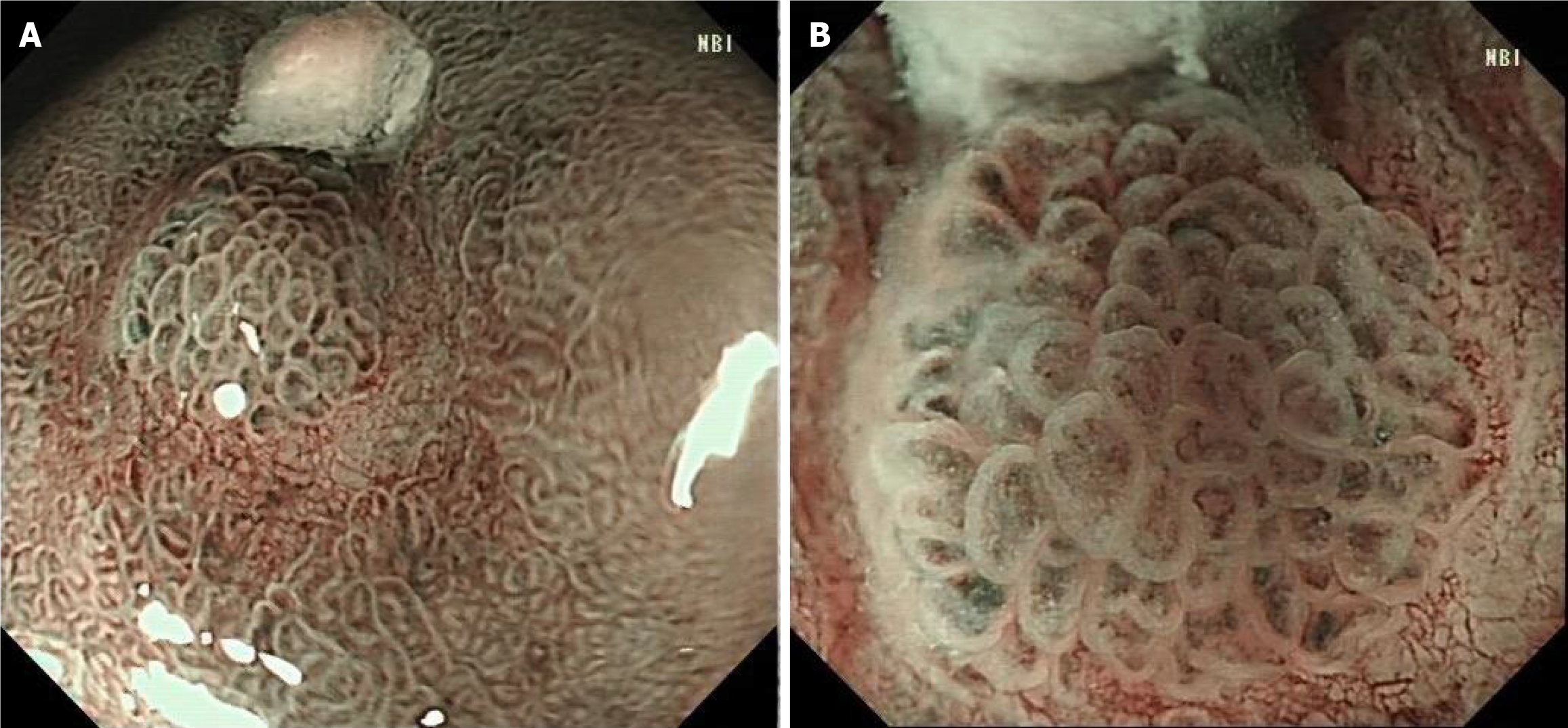
Figure 3 Narrow-band imaging endoscopy.
A: Under narrow-band imaging endoscopy, the lesions were dark tea-coloured and the boundary was clear; B: Under magnifying endoscopy, the micro glandular structure on the surface of the lesions was, of different sizes, and the microvessels were slightly tortuous and expanded, forming a bright boundary with the periphery.
- Citation: Xu YW, Song Y, Tian J, Zhang BC, Yang YS, Wang J. Clinical pathological characteristics of “crawling-type” gastric adenocarcinoma cancer: A case report. World J Gastrointest Oncol 2024; 16(4): 1660-1667
- URL: https://www.wjgnet.com/1948-5204/full/v16/i4/1660.htm
- DOI: https://dx.doi.org/10.4251/wjgo.v16.i4.1660


