Copyright
©The Author(s) 2022.
World J Gastrointest Oncol. Dec 15, 2022; 14(12): 2415-2421
Published online Dec 15, 2022. doi: 10.4251/wjgo.v14.i12.2415
Published online Dec 15, 2022. doi: 10.4251/wjgo.v14.i12.2415
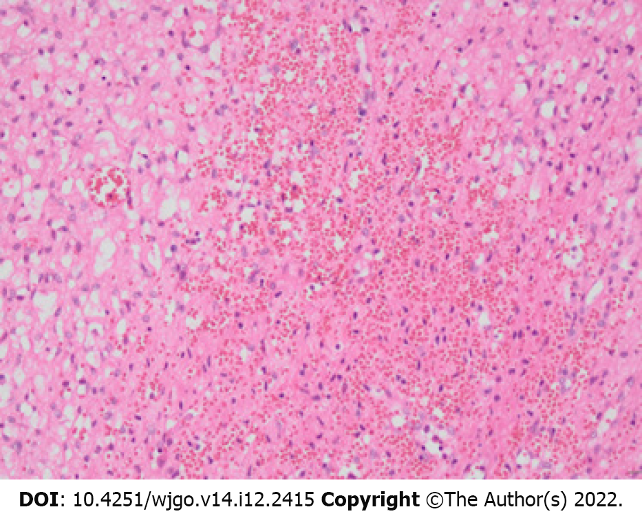
Figure 3 Pathology map (200 ×, hematoxylin-eosin staining).
The tumor was mainly composed of capillaries and eosinophilic, vacuole-containing stromal cells. The cell morphology was mild, the nuclei were small and uniform, and division was rare. The blood vessels were full of blood, with some blood spilling out from the blood vessels.
- Citation: Li DF, Guo XJ, Song SP, Li HB. Rare massive hepatic hemangioblastoma: A case report. World J Gastrointest Oncol 2022; 14(12): 2415-2421
- URL: https://www.wjgnet.com/1948-5204/full/v14/i12/2415.htm
- DOI: https://dx.doi.org/10.4251/wjgo.v14.i12.2415


