
















©The Author(s) 2022.
World J Gastrointest Oncol. Nov 15, 2022; 14(11): 2253-2265
Published online Nov 15, 2022. doi: 10.4251/wjgo.v14.i11.2253
Published online Nov 15, 2022. doi: 10.4251/wjgo.v14.i11.2253
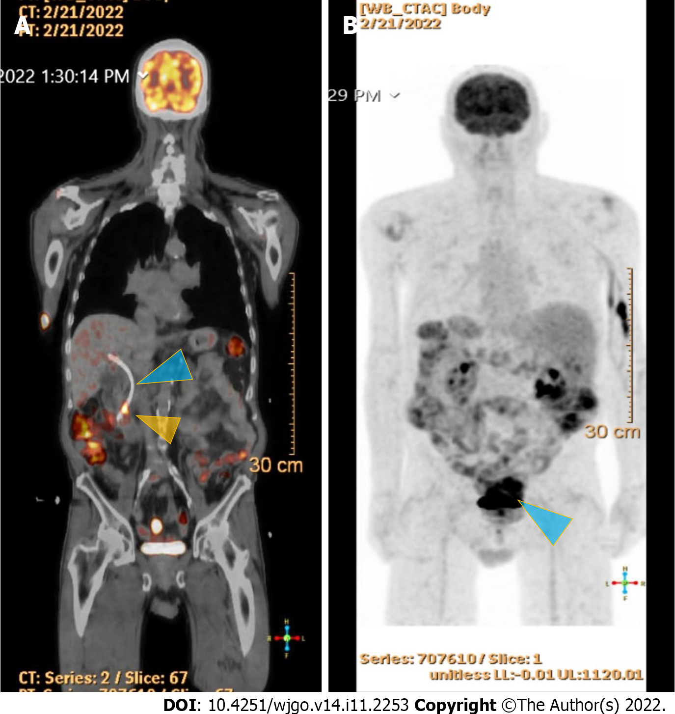
Figure 4 18F-Flurodeoxyglucose positron emission tomography scan studies.
A: Shows an end-obiliary stent in the region of the papilla (blue triangle) and a 1.7 cm ampullary mass with intense fluorodeoxyglucose (FDG) avidity(yellow triangle); B: Shows an oval-shaped, well-defined, FDG-avid lesion measuring approximately 6 cm × 3 cm with a small, punctate area of calcification was present in the lesion located deep in the pelvis along the posterior margin of small bowel loops (blue triangle) with intense FDG avidity.
- Citation: Matli VVK, Zibari GB, Wellman G, Ramadas P, Pandit S, Morris J. A rare synchrony of adenocarcinoma of the ampulla with an ileal gastrointestinal stromal tumor: A case report. World J Gastrointest Oncol 2022; 14(11): 2253-2265
- URL: https://www.wjgnet.com/1948-5204/full/v14/i11/2253.htm
- DOI: https://dx.doi.org/10.4251/wjgo.v14.i11.2253