Copyright
©The Author(s) 2021.
World J Gastrointest Oncol. Sep 15, 2021; 13(9): 1017-1028
Published online Sep 15, 2021. doi: 10.4251/wjgo.v13.i9.1017
Published online Sep 15, 2021. doi: 10.4251/wjgo.v13.i9.1017
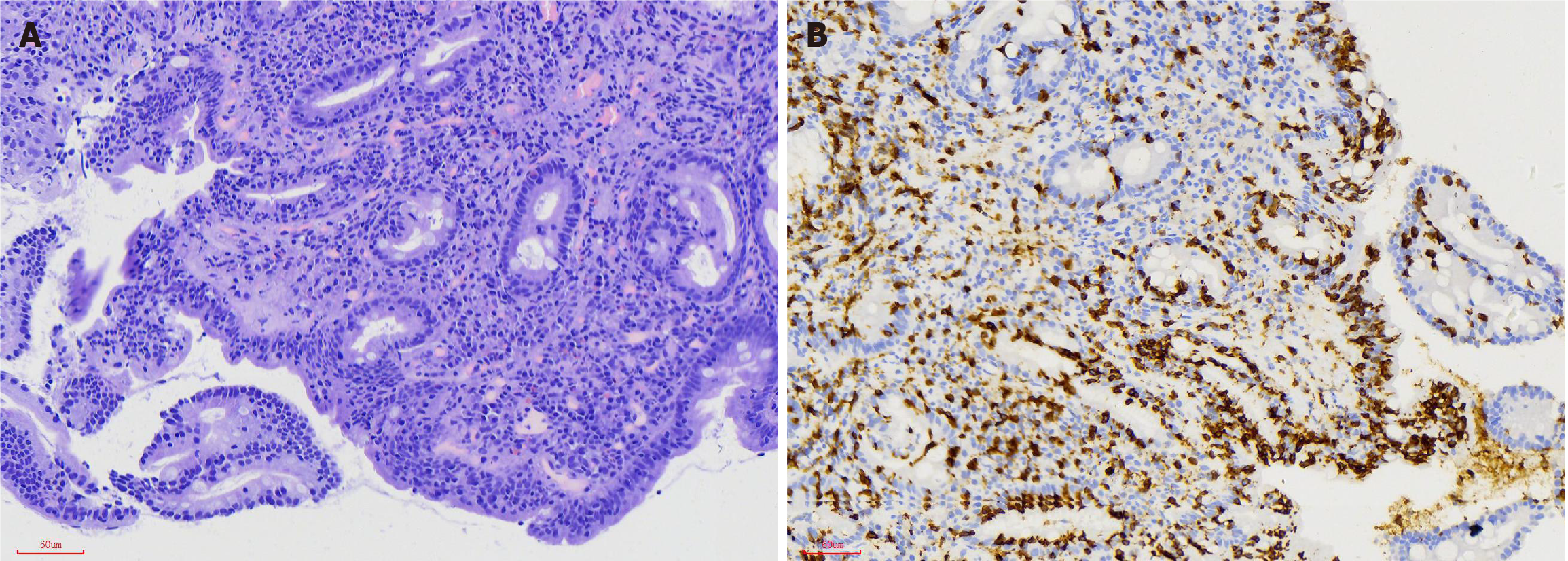
Figure 2 Histologic findings in celiac disease.
A: The specimen shows total villus atrophy and increased intraepithelial lymphocytes (IELs), 60/100 epithelial cells, and crypt hyperplasia (hematoxylin and eosin; original magnification 200 ×); B: CD3 immunohistochemistry (200 ×).
- Citation: Wang M, Yu M, Kong WJ, Cui M, Gao F. Association between intestinal neoplasms and celiac disease: A review. World J Gastrointest Oncol 2021; 13(9): 1017-1028
- URL: https://www.wjgnet.com/1948-5204/full/v13/i9/1017.htm
- DOI: https://dx.doi.org/10.4251/wjgo.v13.i9.1017


