Copyright
©The Author(s) 2021.
World J Gastrointest Oncol. Apr 15, 2021; 13(4): 305-311
Published online Apr 15, 2021. doi: 10.4251/wjgo.v13.i4.305
Published online Apr 15, 2021. doi: 10.4251/wjgo.v13.i4.305
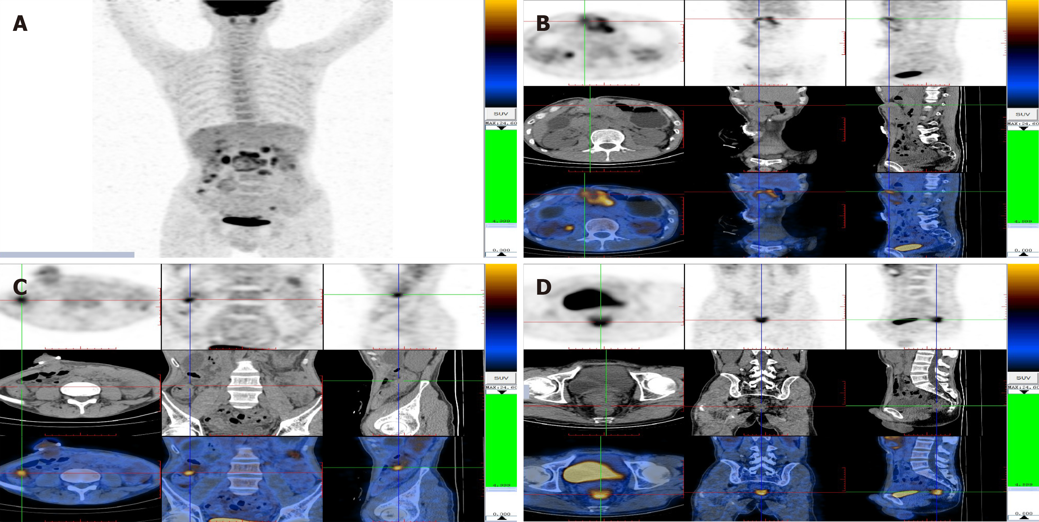
Figure 2 Images from abdominal positron emission tomography-computed tomography scan.
A: There were multiple lesions with increased glucose metabolism in the abdominal cavity; B: The large soft tissue mass adjacent to the transversostomy had high levels of glucose metabolism; C: The ascending colon had high levels of glucose metabolism; D: Multiple nodules were present in the excavatio rectovesicalis with high levels of glucose metabolism.
- Citation: Xiao L, Sun L, Zhao K, Pan YS. Crohn’s disease with infliximab treatment complicated by rapidly progressing colorectal cancer: A case report. World J Gastrointest Oncol 2021; 13(4): 305-311
- URL: https://www.wjgnet.com/1948-5204/full/v13/i4/305.htm
- DOI: https://dx.doi.org/10.4251/wjgo.v13.i4.305


