Copyright
©The Author(s) 2021.
World J Gastrointest Oncol. Oct 15, 2021; 13(10): 1492-1505
Published online Oct 15, 2021. doi: 10.4251/wjgo.v13.i10.1492
Published online Oct 15, 2021. doi: 10.4251/wjgo.v13.i10.1492
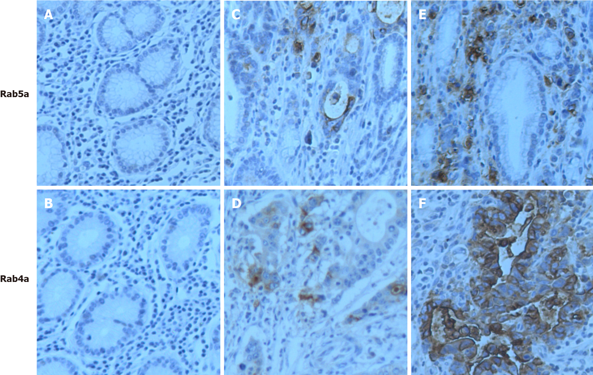
Figure 1 Microscopic images of immunohistochemical staining for Rab4a or Rab5a in gastric tissues.
A and B: Negative (–) in normal tissue sections; C and D: Weak positive (+) in paracancerous tissue sections; E: Strong positive (+++); F; Moderate positive (++) in gastric cancer tissue sections. Original magnification: 200 ×.
- Citation: Cao GJ, Wang D, Zeng ZP, Wang GX, Hu CJ, Xing ZF. Direct interaction between Rab5a and Rab4a enhanced epidermal growth factor-stimulated proliferation of gastric cancer cells. World J Gastrointest Oncol 2021; 13(10): 1492-1505
- URL: https://www.wjgnet.com/1948-5204/full/v13/i10/1492.htm
- DOI: https://dx.doi.org/10.4251/wjgo.v13.i10.1492


