Copyright
©The Author(s) 2020.
World J Gastrointest Oncol. Sep 15, 2020; 12(9): 1031-1043
Published online Sep 15, 2020. doi: 10.4251/wjgo.v12.i9.1031
Published online Sep 15, 2020. doi: 10.4251/wjgo.v12.i9.1031
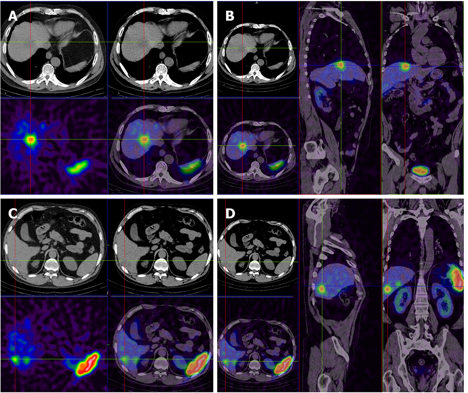
Figure 2 There are multiple low-density lesions in the liver with nonuniform radioactive distribution.
A and B: The size of the largest lesion is 2.3 cm × 2.8 cm, and the computed tomography value is 36 HU; C and D: Smaller lesion.
- Citation: Wang HH, Liu ZC, Zhang G, Li LH, Li L, Meng QB, Wang PJ, Shen DQ, Dang XW. Clinical characteristics and outcome of primary hepatic neuroendocrine tumors after comprehensive therapy. World J Gastrointest Oncol 2020; 12(9): 1031-1043
- URL: https://www.wjgnet.com/1948-5204/full/v12/i9/1031.htm
- DOI: https://dx.doi.org/10.4251/wjgo.v12.i9.1031


