Copyright
©The Author(s) 2018.
World J Gastrointest Oncol. Jan 15, 2018; 10(1): 56-61
Published online Jan 15, 2018. doi: 10.4251/wjgo.v10.i1.56
Published online Jan 15, 2018. doi: 10.4251/wjgo.v10.i1.56
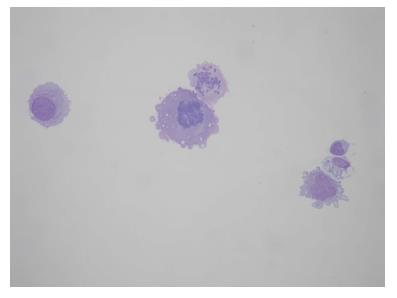
Figure 3 Patient No.
1. Four atypical cells, one lymphocyte and one macrophages next to the lymphocyte. Atypical cells are isolated, two of those show mitotic activity. The size of atypical cells and lymphocyte could be compared (Hemacolor 40 ×).
- Citation: Kountourakis P, Papamichael D, Haralambous H, Michael M, Nakos G, Lazaridou S, Fotiou E, Vassiliou V, Andreopoulos D. Leptomeningeal metastases originated from esophagogastric junction/gastric cancer: A brief report of two cases. World J Gastrointest Oncol 2018; 10(1): 56-61
- URL: https://www.wjgnet.com/1948-5204/full/v10/i1/56.htm
- DOI: https://dx.doi.org/10.4251/wjgo.v10.i1.56


