Copyright
©2009 Baishideng.
World J Gastrointest Oncol. Oct 15, 2009; 1(1): 55-61
Published online Oct 15, 2009. doi: 10.4251/wjgo.v1.i1.55
Published online Oct 15, 2009. doi: 10.4251/wjgo.v1.i1.55
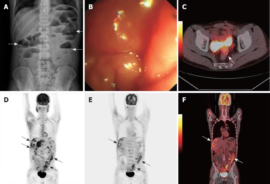
Figure 2 Extrapelvic metastases and perirectal recurrence.
Plain abdominal radiograph, ES and PET/CT images obtained 30 mo after rectal cancer resection in a 41-year-old man. A, B: Plain abdominal radiograph and colonoscopy revealed the obstruction at the anastomotic site (arrows); C-F: PET/CT showed perirectal recurrence, peritoneal carcinomatosis and liver metastases (arrows).
- Citation: Sun L, Guan YS, Pan WM, Luo ZM, Wei JH, Zhao L, Wu H. Clinical value of 18F-FDG PET/CT in assessing suspicious relapse after rectal cancer resection. World J Gastrointest Oncol 2009; 1(1): 55-61
- URL: https://www.wjgnet.com/1948-5204/full/v1/i1/55.htm
- DOI: https://dx.doi.org/10.4251/wjgo.v1.i1.55


