Copyright
©The Author(s) 2016.
World J Gastrointest Oncol. Oct 15, 2016; 8(10): 751-756
Published online Oct 15, 2016. doi: 10.4251/wjgo.v8.i10.751
Published online Oct 15, 2016. doi: 10.4251/wjgo.v8.i10.751
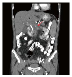
Figure 1 Coronal plain computerized tomography.
Locally advanced pancreatic malignant mass of 45 mm in diameter surrounded and narrowed superior mesenteric artery (arrow).
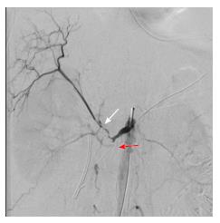
Figure 2 Conventional angiography of superior mesenteric artery immediately after irreversible electroporation.
Angiography revealed that there is occlusion in superior mesenteric artery (red arrow) and also occlusion in hepatic artery (white arrow) originated from superior mesenteric artery.
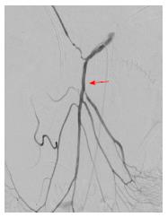
Figure 3 Conventional angiography of superior mesenteric artery after stent placement.
After the stent placement superior mesenteric artery re-canalized and intestinal blood flow was maintained (arrow shows the re-canalized superior mesenteric artery).
- Citation: Ekici Y, Tezcaner T, Aydın HO, Boyvat F, Moray G. Arterial complication of irreversible electroporation procedure for locally advanced pancreatic cancer. World J Gastrointest Oncol 2016; 8(10): 751-756
- URL: https://www.wjgnet.com/1948-5204/full/v8/i10/751.htm
- DOI: https://dx.doi.org/10.4251/wjgo.v8.i10.751


