Copyright
©2014 Baishideng Publishing Group Inc.
World J Gastrointest Oncol. Jun 15, 2014; 6(6): 170-176
Published online Jun 15, 2014. doi: 10.4251/wjgo.v6.i6.170
Published online Jun 15, 2014. doi: 10.4251/wjgo.v6.i6.170
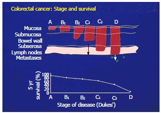
Figure 1 Correlating the extent of local tumor progression with survival in colorectal cancer.
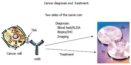
Figure 2 Depicts the ideal monoclonal antibody that can define the presence of a tumor associated antigen for diagnosis by Immunohistochemistry and then when delivered IV, can hunt and destroy the tumor which contains the diagnostic marker.
IHC: Immunohistochemistry; TAA: Tumor associated antigen; ELISA: Enzyme-linked immunosorbent assay.
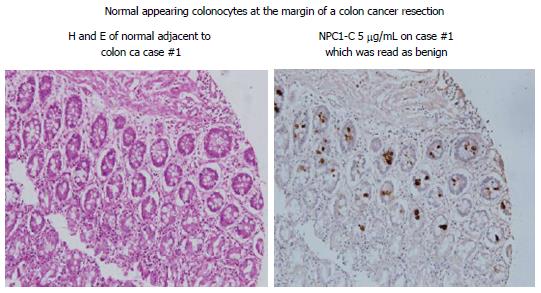
Figure 3 Reveals expression of tumor antigen in those colonocytes adjacent to the malignant lesion where the colonocytes appear normal by H and E.
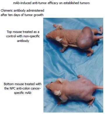
Figure 4 Animal models (nude mice) growing human malignancy to compare untreated and treated animals.
The upper model received a control mAb while the lower animal model having had a much smaller tumor mass at 10 d received mAb NPC-1.
- Citation: Arlen M, Arlen P, Coppa G, Crawford J, Wang X, Saric O, Dubeykovskiy A, Molmenti E. Monoclonal antibodies that target the immunogenic proteins expressed in colorectal cancer. World J Gastrointest Oncol 2014; 6(6): 170-176
- URL: https://www.wjgnet.com/1948-5204/full/v6/i6/170.htm
- DOI: https://dx.doi.org/10.4251/wjgo.v6.i6.170


