
















©The Author(s) 2016.
World J Gastrointest Endosc. Oct 16, 2016; 8(18): 669-673
Published online Oct 16, 2016. doi: 10.4253/wjge.v8.i18.669
Published online Oct 16, 2016. doi: 10.4253/wjge.v8.i18.669
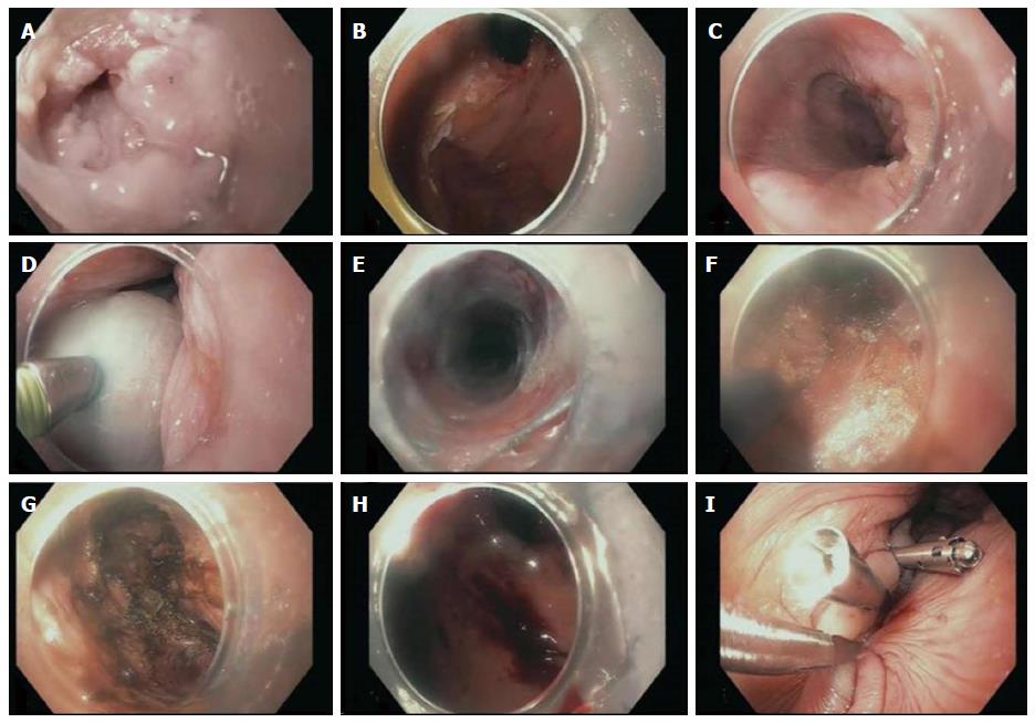
Figure 3 Endoscopic pictures taken from the repeat per oral endoscopic myotomy showing: sigmoid esophagus (A), cardia before myotomy (B), mucosa of previous dissection site (C), Mucosal bleb (D), submucosal tunneling (E), initial myotomy (F), completed myotomy (G), cardia after submucosal tunnneling (H), closure of mucosotomy (I).
- Citation: Wehbeh AN, Mekaroonkamol P, Cai Q. Same site submucosal tunneling for a repeat per oral endoscopic myotomy: A safe and feasible option. World J Gastrointest Endosc 2016; 8(18): 669-673
- URL: https://www.wjgnet.com/1948-5190/full/v8/i18/669.htm
- DOI: https://dx.doi.org/10.4253/wjge.v8.i18.669