
















©2013 Baishideng Publishing Group Co.
World J Gastrointest Endosc. Nov 16, 2013; 5(11): 540-550
Published online Nov 16, 2013. doi: 10.4253/wjge.v5.i11.540
Published online Nov 16, 2013. doi: 10.4253/wjge.v5.i11.540
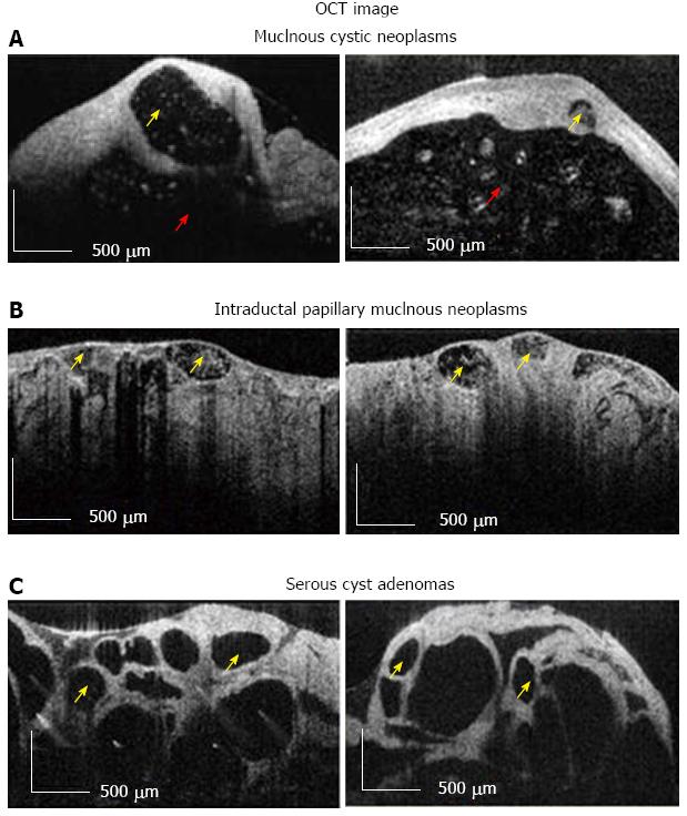
Figure 8 Optical coherence tomography image.
A, B: Diagnostic criteria for high-risk (i.e., Mucinous Cystic Neoplasms, Intraductal Papillary Mucinous Neoplasms); C: Low risk (i.e., Serous Cysts Adenomas) pancreatic cysts. Multiple small cysts are marked with yellow arrow, while surrounded main cystic cavity is marked with red arrow. Scale bar = 500 μm[69]. OCT: Optical coherence tomography.
- Citation: Mahmud MS, May GR, Kamal MM, Khwaja AS, Sun C, Vitkin A, Yang VX. Imaging pancreatobiliary ductal system with optical coherence tomography: A review. World J Gastrointest Endosc 2013; 5(11): 540-550
- URL: https://www.wjgnet.com/1948-5190/full/v5/i11/540.htm
- DOI: https://dx.doi.org/10.4253/wjge.v5.i11.540