Copyright
©The Author(s) 2016.
World J Hepatol. Nov 28, 2016; 8(33): 1419-1441
Published online Nov 28, 2016. doi: 10.4254/wjh.v8.i33.1419
Published online Nov 28, 2016. doi: 10.4254/wjh.v8.i33.1419
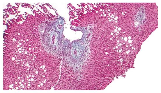
Figure 4 Histology of primary sclerosing cholangitis.
Trichrome × 40, liver biopsy. Low power view demonstrating a focal lesion typical for primary sclerosing cholangitis. Periductular layered fibrosis (featuring “onion skin” pattern) is found with edema and inflammation around the interlobular bile ducts in the center of the field[9].
- Citation: Huang YQ. Recent advances in the diagnosis and treatment of primary biliary cholangitis. World J Hepatol 2016; 8(33): 1419-1441
- URL: https://www.wjgnet.com/1948-5182/full/v8/i33/1419.htm
- DOI: https://dx.doi.org/10.4254/wjh.v8.i33.1419


