Copyright
©The Author(s) 2016.
World J Hepatol. Nov 28, 2016; 8(33): 1419-1441
Published online Nov 28, 2016. doi: 10.4254/wjh.v8.i33.1419
Published online Nov 28, 2016. doi: 10.4254/wjh.v8.i33.1419
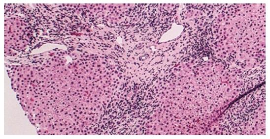
Figure 1 Histology of primary biliary cholangitis (hematoxylin and eosin staining; × 200 liver biopsy).
An absence/paucity of bile ducts is seen with focal chronic inflammation in a portal area consistent with late-stage primary biliary cholangitis[9].
- Citation: Huang YQ. Recent advances in the diagnosis and treatment of primary biliary cholangitis. World J Hepatol 2016; 8(33): 1419-1441
- URL: https://www.wjgnet.com/1948-5182/full/v8/i33/1419.htm
- DOI: https://dx.doi.org/10.4254/wjh.v8.i33.1419


