Copyright
©The Author(s) 2015.
World J Hepatol. Nov 18, 2015; 7(26): 2696-2702
Published online Nov 18, 2015. doi: 10.4254/wjh.v7.i26.2696
Published online Nov 18, 2015. doi: 10.4254/wjh.v7.i26.2696
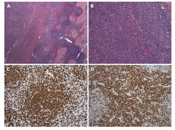
Figure 2 Histopathological findings of case 1.
A and B: Tumoral nodule containing lymphoid proliferation characterized by reactive lymphoid follicles and interfollicular plasma cells within liver parenchyma; C: Numerous CD20 positive B cells mostly confined to follicles; D: Numerous interfollicular polyclonal CD138 and MUM-1 positive plasma cells.
- Citation: Kwon YK, Jha RC, Etesami K, Fishbein TM, Ozdemirli M, Desai CS. Pseudolymphoma (reactive lymphoid hyperplasia) of the liver: A clinical challenge. World J Hepatol 2015; 7(26): 2696-2702
- URL: https://www.wjgnet.com/1948-5182/full/v7/i26/2696.htm
- DOI: https://dx.doi.org/10.4254/wjh.v7.i26.2696


