Copyright
©The Author(s) 2015.
World J Hepatol. Feb 27, 2015; 7(2): 276-284
Published online Feb 27, 2015. doi: 10.4254/wjh.v7.i2.276
Published online Feb 27, 2015. doi: 10.4254/wjh.v7.i2.276
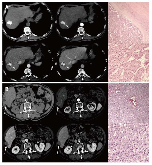
Figure 4 Multidetector CT scans demonstrating a hypervascular area representing viable hepatocellular carcinoma next to a previously lipiodolized nodule, graded as moderately differentiated on histopathology (HE stain, magnification × 40 and × 200) (A); Multidetector CT scans of the same patients 1 year after LT demonstrating a hypervascular intrahepatic recurrent hepatocellular carcinoma, which was graded as poorly differentiated on histopathology after resection (HE stain, magnification × 40 and × 200) (B).
- Citation: Pecchi A, Besutti G, Santis MD, Giovane CD, Nosseir S, Tarantino G, Benedetto FD, Torricelli P. Post-transplantation hepatocellular carcinoma recurrence: Patterns and relation between vascularity and differentiation degree. World J Hepatol 2015; 7(2): 276-284
- URL: https://www.wjgnet.com/1948-5182/full/v7/i2/276.htm
- DOI: https://dx.doi.org/10.4254/wjh.v7.i2.276


