Copyright
©The Author(s) 2015.
World J Hepatol. Feb 27, 2015; 7(2): 276-284
Published online Feb 27, 2015. doi: 10.4254/wjh.v7.i2.276
Published online Feb 27, 2015. doi: 10.4254/wjh.v7.i2.276
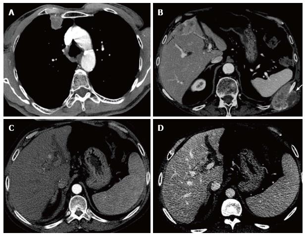
Figure 2 Representative multidetector CT images of different recurrence patterns based on the location at the moment of the first recurrence: (A) Extrahepatic recurrence presenting with a solitary pleural-based lung nodule; (B) Both intrahepatic and extrahepatic recurrence presenting with bone lesions and an intrahepatic lesion in segments IVb and V; (C and D) Intrahepatic recurrence presenting with a solitary hypervascular nodule in segment IV.
- Citation: Pecchi A, Besutti G, Santis MD, Giovane CD, Nosseir S, Tarantino G, Benedetto FD, Torricelli P. Post-transplantation hepatocellular carcinoma recurrence: Patterns and relation between vascularity and differentiation degree. World J Hepatol 2015; 7(2): 276-284
- URL: https://www.wjgnet.com/1948-5182/full/v7/i2/276.htm
- DOI: https://dx.doi.org/10.4254/wjh.v7.i2.276


