Copyright
©The Author(s) 2015.
World J Hepatol. Feb 27, 2015; 7(2): 276-284
Published online Feb 27, 2015. doi: 10.4254/wjh.v7.i2.276
Published online Feb 27, 2015. doi: 10.4254/wjh.v7.i2.276
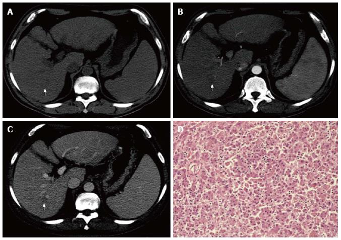
Figure 1 Pre-contrast (A), hepatic arterial (B) and hepatic portal venous (C) phases multidetector CT scans demonstrate a hypovascular hepatocellular carcinoma (arrows), with no hypervascularization during the arterial phase (wash-in).
Pathologically (HE stain, × 100) (D), it was classified as well-differentiated (Grade 1).
- Citation: Pecchi A, Besutti G, Santis MD, Giovane CD, Nosseir S, Tarantino G, Benedetto FD, Torricelli P. Post-transplantation hepatocellular carcinoma recurrence: Patterns and relation between vascularity and differentiation degree. World J Hepatol 2015; 7(2): 276-284
- URL: https://www.wjgnet.com/1948-5182/full/v7/i2/276.htm
- DOI: https://dx.doi.org/10.4254/wjh.v7.i2.276


