Copyright
©The Author(s) 2015.
World J Hepatol. Jun 28, 2015; 7(12): 1632-1651
Published online Jun 28, 2015. doi: 10.4254/wjh.v7.i12.1632
Published online Jun 28, 2015. doi: 10.4254/wjh.v7.i12.1632
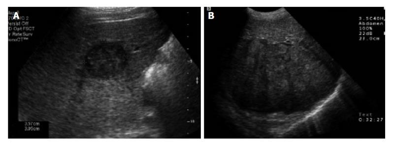
Figure 1 Abdominal ultrasound of the liver.
A: Transverse sonogram shows a small, 3 cm, hypoechoic mass in the right lobe of the liver; B: Transverse sonogram shows a heterogeneous large mass in the right lobe of the liver[47].
- Citation: Attwa MH, El-Etreby SA. Guide for diagnosis and treatment of hepatocellular carcinoma. World J Hepatol 2015; 7(12): 1632-1651
- URL: https://www.wjgnet.com/1948-5182/full/v7/i12/1632.htm
- DOI: https://dx.doi.org/10.4254/wjh.v7.i12.1632


