Copyright
©2014 Baishideng Publishing Group Inc.
World J Hepatol. Aug 27, 2014; 6(8): 580-595
Published online Aug 27, 2014. doi: 10.4254/wjh.v6.i8.580
Published online Aug 27, 2014. doi: 10.4254/wjh.v6.i8.580
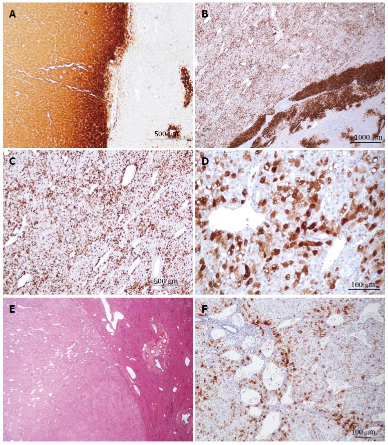
Figure 17 β-hepatocellular adenoma different types of glutamine synthase immunostaining.
A: Same patient as Figure 16C, D. Strong and diffuse expression of glutamine synthase (GS) in hepatocellular adenoma (HCA) (left), contrasting with non tumoral liver (positivity in only few pericentrolobular hepatocytes). B-D: Same patient as Figure 16 E, F. Strong, heterogeneous (patchy) positivity of GS seen at different magnification, with a reinforcement rim at the periphery of the HCA (the rim positivity has no pathological significance). E, F: Woman born in 1986; abnormal liver function tests. Imaging: one nodule 9 cm, HCA. Left hepatectomy 2007. E: HE - no specific abnormalities. The presence of numerous vessels is, however, intriguing in this young patient. F: Patchy positivity of GS from mild to strong.
- Citation: Sempoux C, Balabaud C, Bioulac-Sage P. Pictures of focal nodular hyperplasia and hepatocellular adenomas. World J Hepatol 2014; 6(8): 580-595
- URL: https://www.wjgnet.com/1948-5182/full/v6/i8/580.htm
- DOI: https://dx.doi.org/10.4254/wjh.v6.i8.580


