Copyright
©2014 Baishideng Publishing Group Inc.
World J Hepatol. Aug 27, 2014; 6(8): 580-595
Published online Aug 27, 2014. doi: 10.4254/wjh.v6.i8.580
Published online Aug 27, 2014. doi: 10.4254/wjh.v6.i8.580
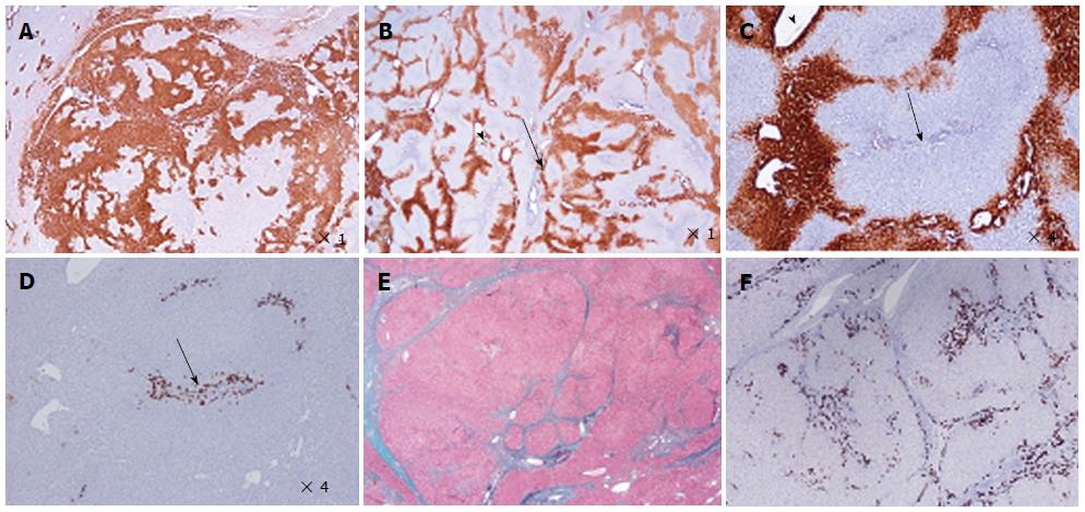
Figure 2 Focal nodular hyperplasia microscopic typical features on Masson’s trichrome, glutamine synthase and CK7 immunostainings.
A: Same patient as Figure 1C. Glutamine synthase (GS) immunostaining: typical aspect of focal nodular hyperplasia (FNH) with large anastomosed positive areas in a “map-like” pattern[11]. B-D: Woman born in 1969; abnormal liver function tests (GGT); ultrasound (US): nodule interpreted as a hemangioma, computed tomography scan and magnetic resonance imaging: FNH 8 cm, segment VIII with minor dilatation of the right biliary tree (compression by the tumor). Left lobectomy + segmentectomy VIII in 2002 (other nodule segment II: 0.5 cm). B, C: GS immunostaining: typical aspect of FNH with anastomosed positive areas often centered by veins (arrowhead) and at distance of fibrous bands (arrows). C and D are from 2 serial sections. D: CK7 immunostaining: ductular reaction at the periphery of fibrous bands. E, F: Same patient as Figure 1C. E: Masson’s trichrome - fibrous bands surrounding benign hepatocytic nodules of different sizes. F: CK7 immunostaining - prominent ductular reaction at the junction between parenchymal nodules and fibrous bands. Typical aspect of FNH.
- Citation: Sempoux C, Balabaud C, Bioulac-Sage P. Pictures of focal nodular hyperplasia and hepatocellular adenomas. World J Hepatol 2014; 6(8): 580-595
- URL: https://www.wjgnet.com/1948-5182/full/v6/i8/580.htm
- DOI: https://dx.doi.org/10.4254/wjh.v6.i8.580


