Copyright
©2010 Baishideng.
World J Hepatol. Jun 27, 2010; 2(6): 239-242
Published online Jun 27, 2010. doi: 10.4254/wjh.v2.i6.239
Published online Jun 27, 2010. doi: 10.4254/wjh.v2.i6.239
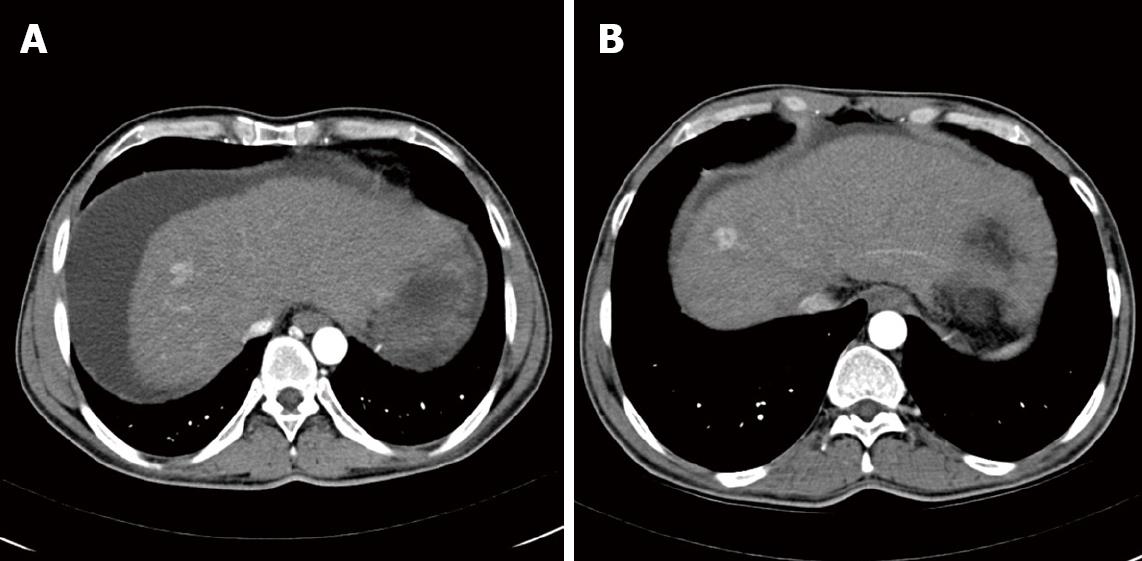
Figure 1 Computed tomography (CT)-scans: Axial IV contrast enhanced CT.
Arterial phase image showing the reference lesion at the indicated time points. Size of this lesion remained stable between 2007 and 2009. A: CT liver November, 2007; B: CT liver December, 2009.
- Citation: Roderburg C, Bubenzer J, Spannbauer M, O ND, Mahnken A, Ludde T, Trautwein C, Tischendorf JJ. Long-term survival of a HCC-patient with severe liver dysfunction treated with sorafenib. World J Hepatol 2010; 2(6): 239-242
- URL: https://www.wjgnet.com/1948-5182/full/v2/i6/239.htm
- DOI: https://dx.doi.org/10.4254/wjh.v2.i6.239


