
















©The Author(s) 2026.
World J Hepatol. Feb 27, 2026; 18(2): 113686
Published online Feb 27, 2026. doi: 10.4254/wjh.v18.i2.113686
Published online Feb 27, 2026. doi: 10.4254/wjh.v18.i2.113686
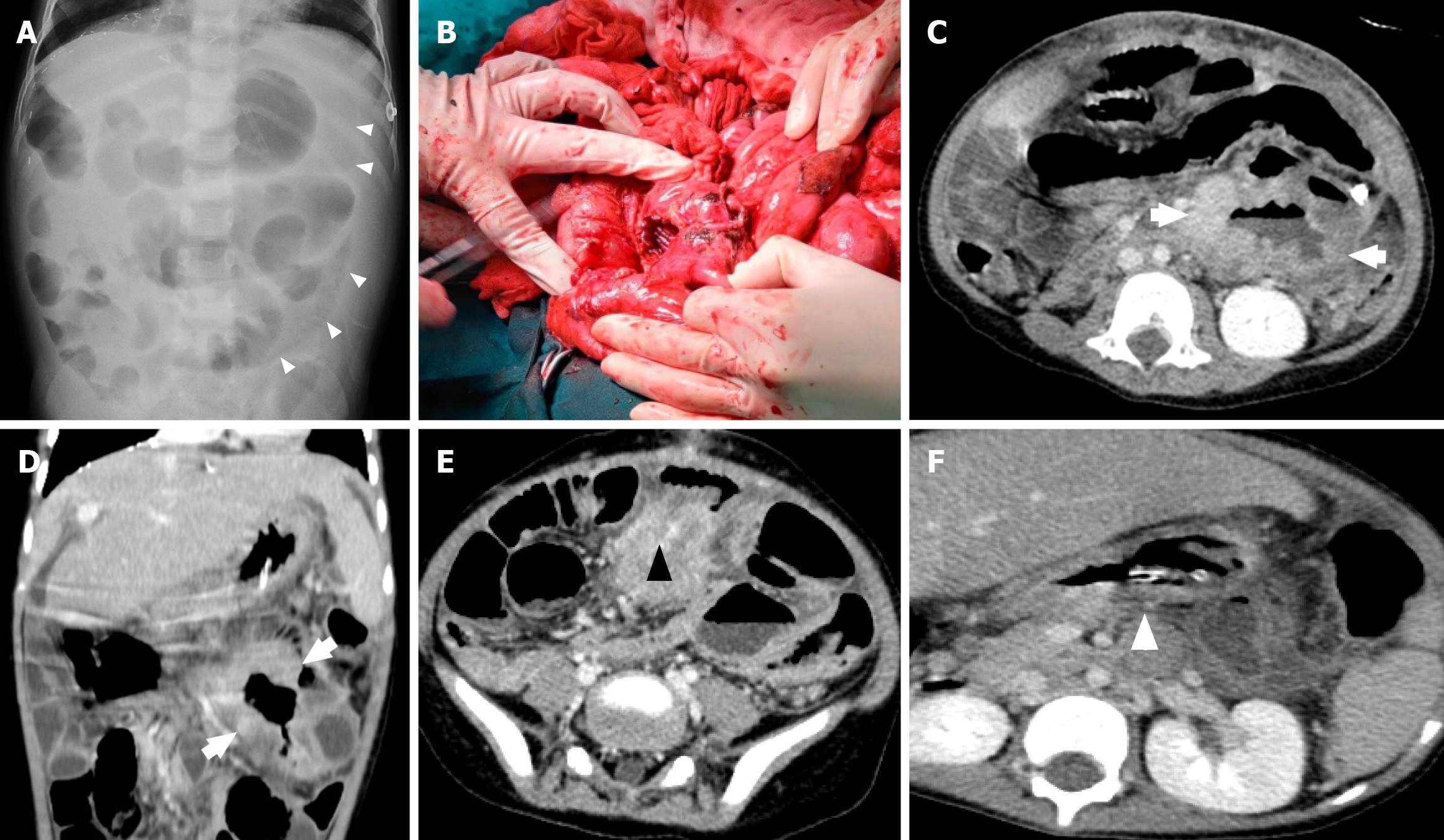
Figure 2 A 4-year-old boy who underwent liver transplantation 3 years ago presented with a subacute fever and abdominal pain.
Supine abdominal radiograph showed free intraperitoneal air (white triangle in Figure 2A) and an infiltrative lesion at the duodenojejunal site, a 6 cm mass with a large perforation site at the duodenojejunal junction. Computed tomography images 7 days after exploratory laparotomy showed segmental, irregular bowel wall thickening and aneurysmal dilation of a jejunal loop in the left upper to mid-abdomen (white arrows), matted mesenteric lymph nodes (black triangle), and an enlarged para-aortic lymph node (white triangle). A: Abdominal X-ray; B: Operative finding; C-F: Computed tomography abdomen (cross section).
- Citation: Onpoaree N, Leelakanok N, Sanpavat A, Sintusek P. Epstein-Barr virus infection in children with liver transplantation. World J Hepatol 2026; 18(2): 113686
- URL: https://www.wjgnet.com/1948-5182/full/v18/i2/113686.htm
- DOI: https://dx.doi.org/10.4254/wjh.v18.i2.113686