Copyright
©The Author(s) 2025.
World J Hepatol. Aug 27, 2025; 17(8): 108443
Published online Aug 27, 2025. doi: 10.4254/wjh.v17.i8.108443
Published online Aug 27, 2025. doi: 10.4254/wjh.v17.i8.108443
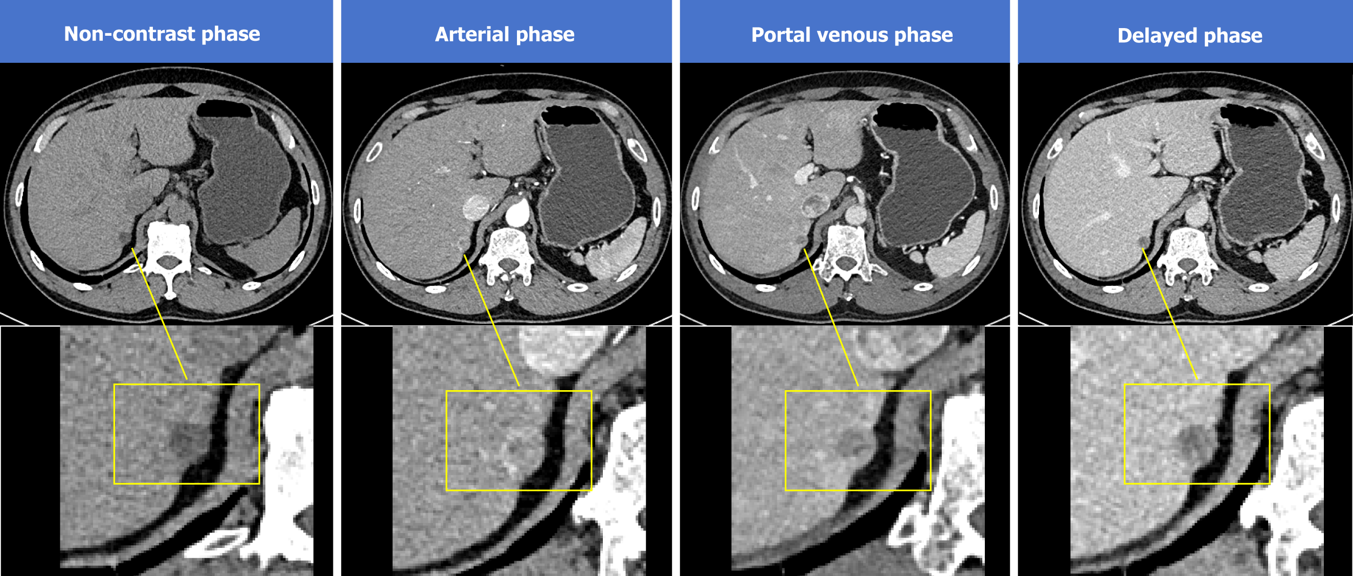
Figure 1 Computed tomography imaging.
A nodular hypodense lesion was observed in segment 7 of the liver, approximately 12 mm in diameter, with well-defined borders. The lesion indicated significant heterogeneous enhancement in the arterial phase, mild attenuation in the portal phase, and marked attenuation in the delayed phase.
- Citation: Qin MQ, Zhao YP, Xie JP. Ectopic adrenal gland in the liver leading to a misdiagnosis of hepatocellular carcinoma: A case report. World J Hepatol 2025; 17(8): 108443
- URL: https://www.wjgnet.com/1948-5182/full/v17/i8/108443.htm
- DOI: https://dx.doi.org/10.4254/wjh.v17.i8.108443


