Copyright
©The Author(s) 2025.
World J Hepatol. Jun 27, 2025; 17(6): 108558
Published online Jun 27, 2025. doi: 10.4254/wjh.v17.i6.108558
Published online Jun 27, 2025. doi: 10.4254/wjh.v17.i6.108558
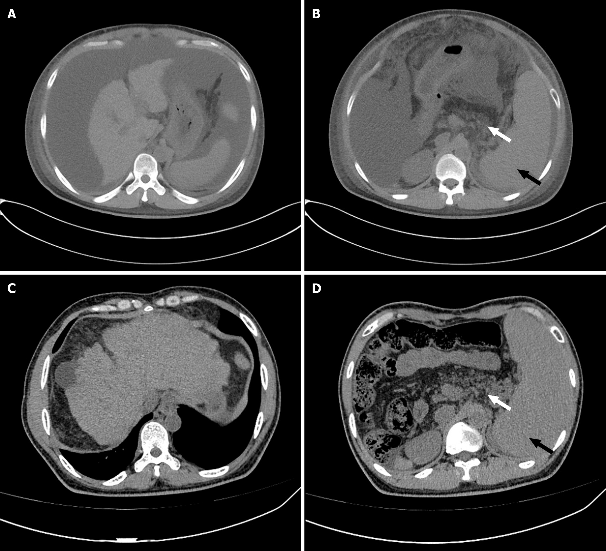
Figure 1 An abdominal computed tomography scan.
A: Liver cirrhosis; B: Splenomegaly; C: Pancreatic fat infiltration; D: A significant accumulation of ascites.
- Citation: Guo HJ. Adult presentation of Shwachman-Diamond syndrome complicated by liver cirrhosis and pancreatic fat infiltration: A case report. World J Hepatol 2025; 17(6): 108558
- URL: https://www.wjgnet.com/1948-5182/full/v17/i6/108558.htm
- DOI: https://dx.doi.org/10.4254/wjh.v17.i6.108558


