Copyright
©The Author(s) 2021.
World J Hepatol. Mar 27, 2021; 13(3): 343-361
Published online Mar 27, 2021. doi: 10.4254/wjh.v13.i3.343
Published online Mar 27, 2021. doi: 10.4254/wjh.v13.i3.343
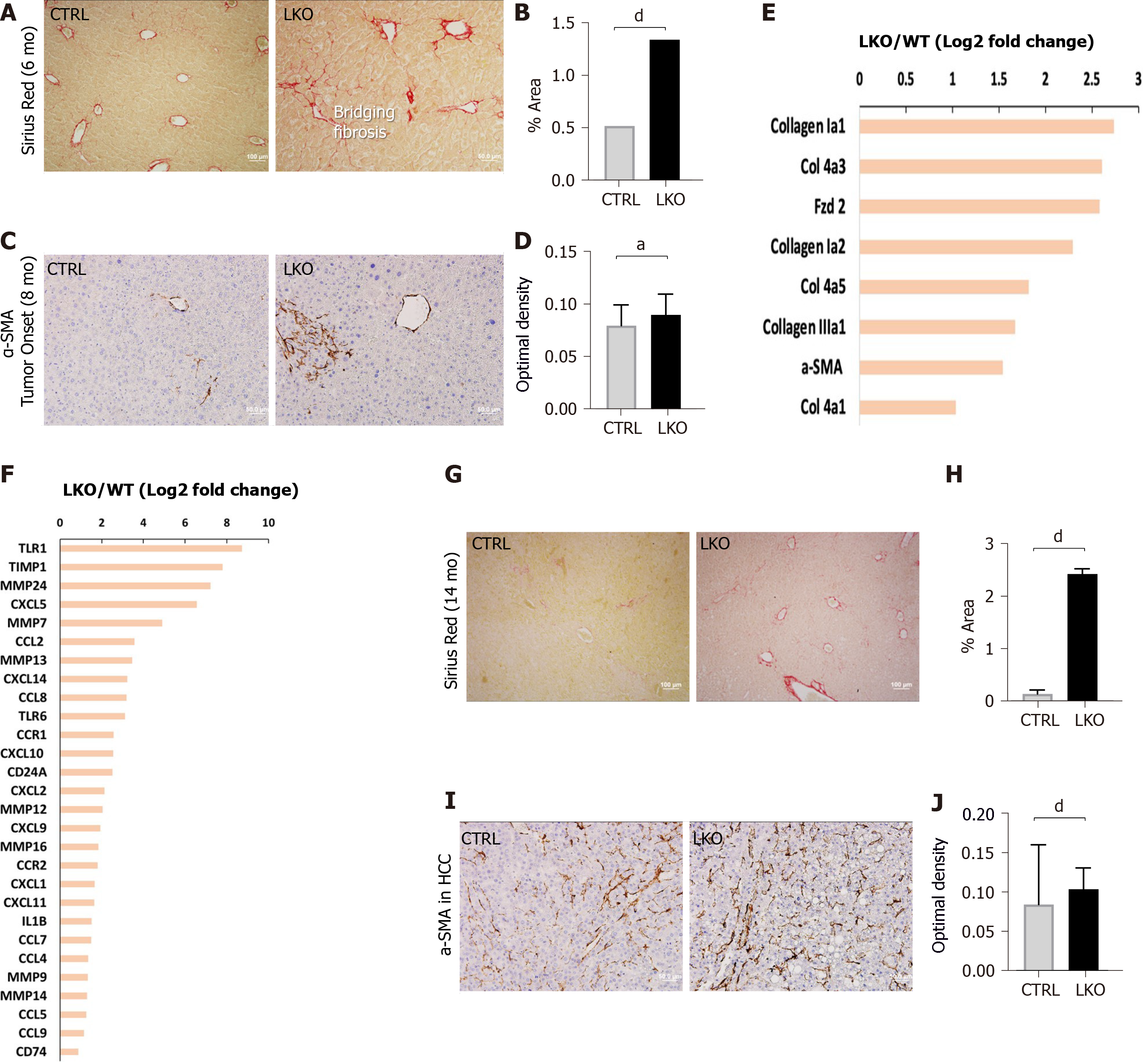
Figure 3 BRUCE deficiency accelerates and increases diethylnitrosamine-induced fibrosis.
A and B: Sirius red staining of control and liver-specific BRUCE KO livers 6 mo post-diethylnitrosamine (DEN) exposure show an increase of Sirius red staining in the LKO livers, verified by quantification to the right; C and D: α-smooth muscle actin (SMA) immunohistochemistry at LKO tumor onset show an increase in α-SMA in LKO livers which is quantified in; E and F: RNA-seq analysis at the time of LKO tumor onset reveal that LKO livers demonstrate key patterns of human hepatic fibrosis, such as increased collagens and increased α-SMA as well as increased inflammation-related markers, such as CCL2; G and H: Sirius red staining of 14 mo post-DEN exposed HCC livers reveal an increase of collagen deposition, which was quantified; I and J: α-SMA immunohistochemistry of HCC livers, including quantification demonstrate an increase of activated hepatic stellate cells in 14 mo DEN-exposed LKO livers. aP < 0.05; dP < 0.001. HCC: Hepatocellular carcinoma; LKO: Liver-specific knockout; α-SMA: α-smooth muscle actin; CTRL: Control.
- Citation: Vilfranc CL, Che LX, Patra KC, Niu L, Olowokure O, Wang J, Shah SA, Du CY. BIR repeat-containing ubiquitin conjugating enzyme (BRUCE) regulation of β-catenin signaling in the progression of drug-induced hepatic fibrosis and carcinogenesis. World J Hepatol 2021; 13(3): 343-361
- URL: https://www.wjgnet.com/1948-5182/full/v13/i3/343.htm
- DOI: https://dx.doi.org/10.4254/wjh.v13.i3.343


