Copyright
©The Author(s) 2021.
World J Hepatol. Feb 27, 2021; 13(2): 261-269
Published online Feb 27, 2021. doi: 10.4254/wjh.v13.i2.261
Published online Feb 27, 2021. doi: 10.4254/wjh.v13.i2.261
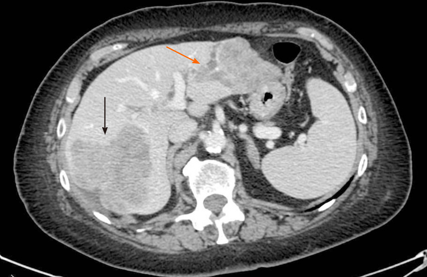
Figure 1 Pre-operative computed tomography-scan.
The lesion occupying the right posterior segments of the liver (black arrow) and two other confluent lesions in the left lobe with intrabiliary growth pattern (orange arrow).
- Citation: Serenari M, Neri J, Marasco G, Larotonda C, Cappelli A, Ravaioli M, Mosconi C, Golfieri R, Cescon M. Two-stage hepatectomy with radioembolization for bilateral colorectal liver metastases: A case report. World J Hepatol 2021; 13(2): 261-269
- URL: https://www.wjgnet.com/1948-5182/full/v13/i2/261.htm
- DOI: https://dx.doi.org/10.4254/wjh.v13.i2.261


