
















©The Author(s) 2020.
World J Hepatol. Dec 27, 2020; 12(12): 1198-1210
Published online Dec 27, 2020. doi: 10.4254/wjh.v12.i12.1198
Published online Dec 27, 2020. doi: 10.4254/wjh.v12.i12.1198
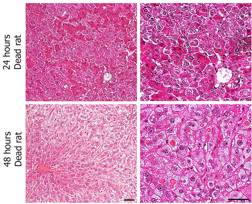
Figure 2 Severity of hepatocyte damage on the onset of liver regeneration.
Histological analysis of representative liver sections harvested within minutes of death and stained with hematoxylin, eosin, and alcian blue from rats that died 24 h and 48 h post-resection. Note the presence of enlarged hepatocytes containing cytoplasmic hyaline inclusions. Scale bar: 50 μmol/L.
- Citation: Moniaux N, Lacaze L, Gothland A, Deshayes A, Samuel D, Faivre J. Cyclin-dependent kinase inhibitors p21 and p27 function as critical regulators of liver regeneration following 90% hepatectomy in the rat. World J Hepatol 2020; 12(12): 1198-1210
- URL: https://www.wjgnet.com/1948-5182/full/v12/i12/1198.htm
- DOI: https://dx.doi.org/10.4254/wjh.v12.i12.1198