Copyright
©2009 Baishideng.
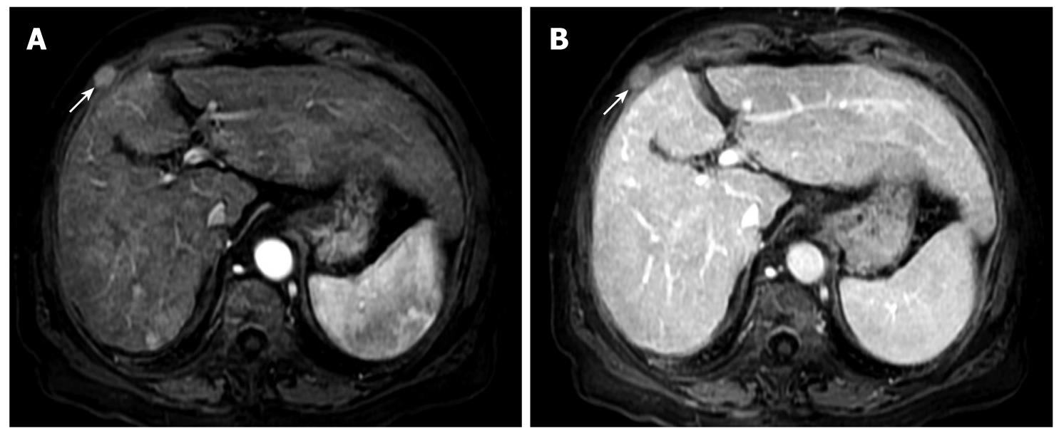
Figure 2 MRI features of HCC seeding following RFA.
A: An arterial phase MRI examination of the liver and an enhancing mass in the intercostal space, indicated by the arrowheads, in the right hemithorax; B: A delayed phase MRI examination that shows the mass in the intercostal space, but this is hypointense compared with the rest of the liver.
- Citation: Cabibbo G, Craxì A. Needle track seeding following percutaneous procedures for hepatocellular carcinoma. World J Hepatol 2009; 1(1): 62-66
- URL: https://www.wjgnet.com/1948-5182/full/v1/i1/62.htm
- DOI: https://dx.doi.org/10.4254/wjh.v1.i1.62


