Copyright
©2009 Baishideng.
World J Hepatol. Oct 31, 2009; 1(1): 103-109
Published online Oct 31, 2009. doi: 10.4254/wjh.v1.i1.103
Published online Oct 31, 2009. doi: 10.4254/wjh.v1.i1.103
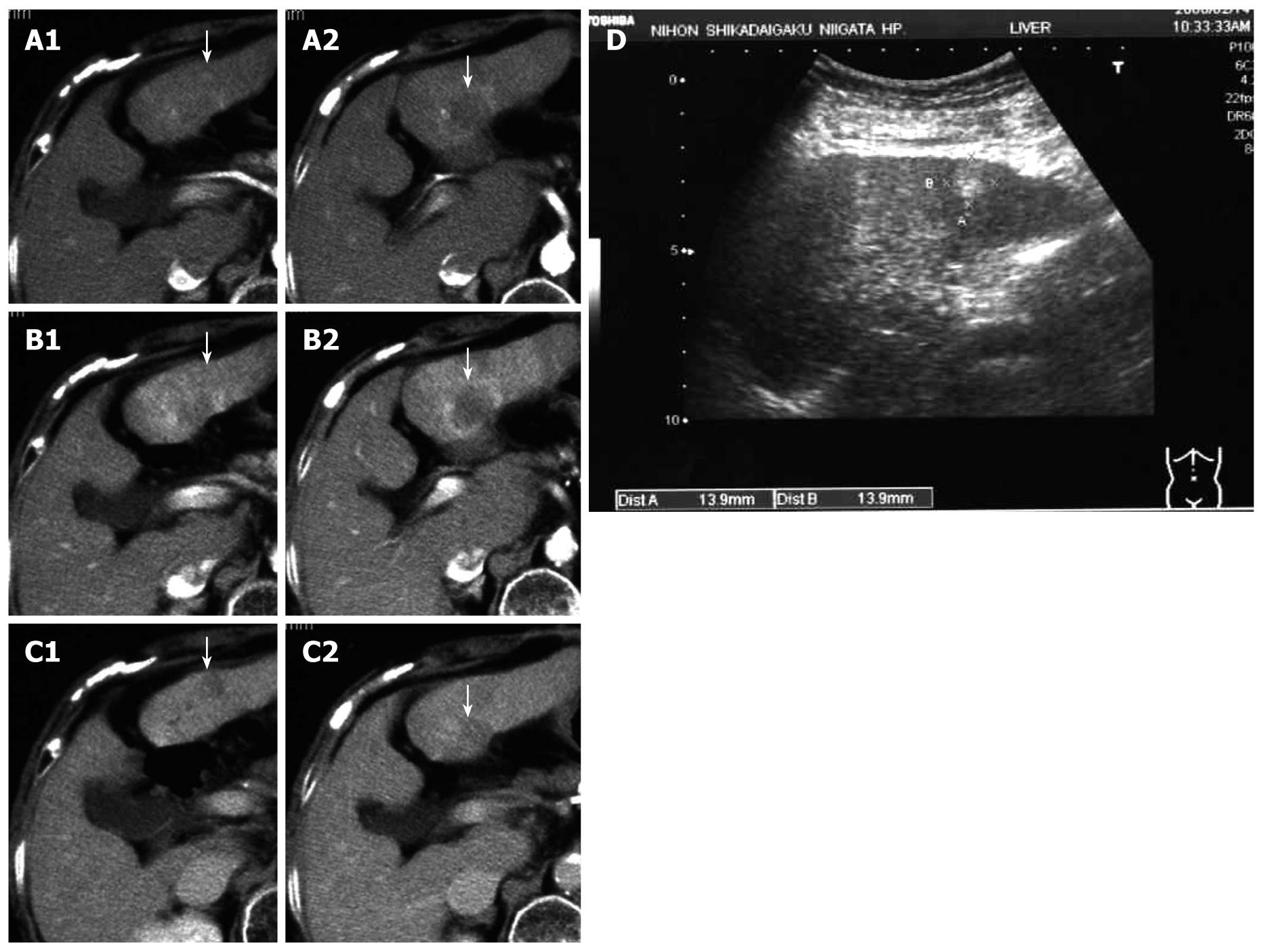
Figure 2 Dynamic computed tomography performed 12 mo after the first operation.
The tumor on the ventral side of S3 appears to be a classic hepatocellular carcinoma and that on the dorsal side of S3 appears to be increased to ~3 cm in diameter. Typical findings including enhancement of the peripheral portion of the tumor in the early (A1) and parenchymal (B2) phases, and the slight and gradual enhancement of the internal portion in the delayed phase were observed (C2). Arrows indicate the tumors (A1, A2 arterial phase; B1, B2 parenchymal phase; C1, C2 delayed phase). Subcutaneous ultrasonography performed 12 mo after the first operation (D); The tumor on the ventral side of S3 is represented by a hyperechoic mass, 14 mm in diameter, a finding characteristic of hepatocellular carcinoma rich in fat. The tumor on the dorsal side of S3 is also represented by a hyperechoic lesion, ~30 mm in diameter, with irregular and unclear margins. Arrows indicate the tumors (D).
- Citation: Watanabe T, Sakata J, Ishikawa T, Shirai Y, Suda T, Hirono H, Hasegawa K, Soga K, Shibasaki K, Saito Y, Umezu H. Synchronous development of HCC and CCC in the same subsegment of the liver in a patient with type C liver cirrhosis. World J Hepatol 2009; 1(1): 103-109
- URL: https://www.wjgnet.com/1948-5182/full/v1/i1/103.htm
- DOI: https://dx.doi.org/10.4254/wjh.v1.i1.103


