Copyright
©2012 Baishideng Publishing Group Co.
World J Hepatol. Nov 27, 2012; 4(11): 322-326
Published online Nov 27, 2012. doi: 10.4254/wjh.v4.i11.322
Published online Nov 27, 2012. doi: 10.4254/wjh.v4.i11.322
Figure 1 Preoperative computed tomography scan revealed a 28 mm tumor in the posterior sector of the right hepatic lobe.
A: Plain computed tomography showed a tumor as a slight low density area; B: The tumor was inhomogeneously enhanced with a ragged border during the early phase; C: The tumor was indistinguishable in the late phase.
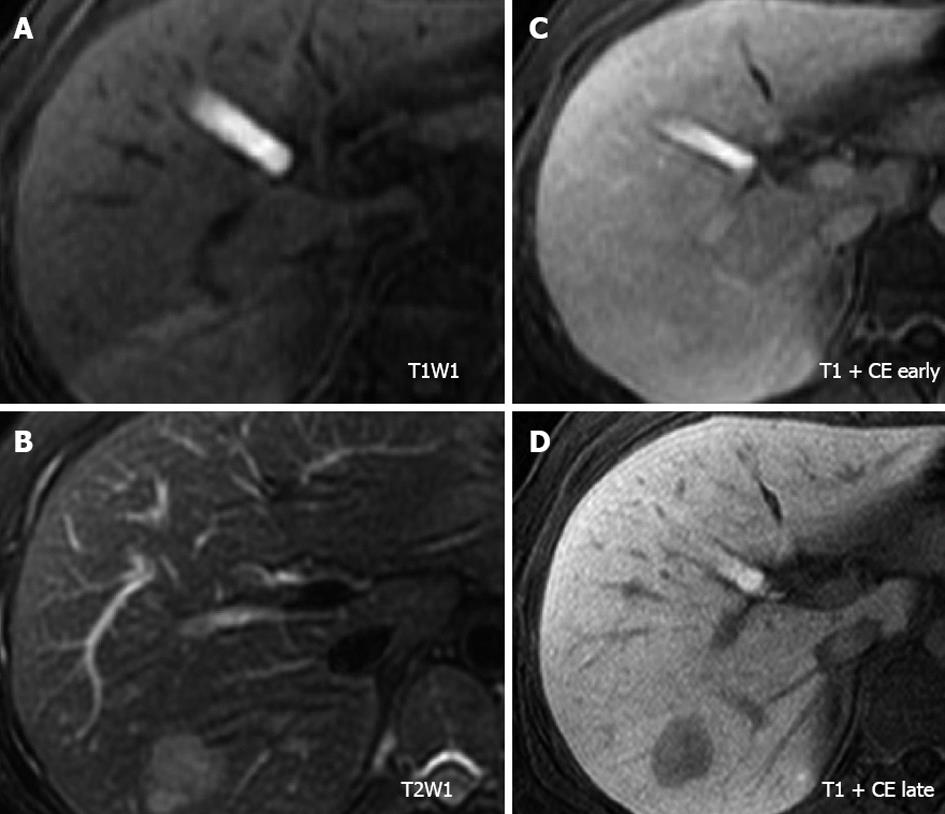
Figure 2 Magnetic resonance imaging of the tumor.
A: The tumor showed an iso-intensity with the surrounding liver parenchyma on T1-weighted imaging; B: The tumor was visualized as a heterogeneous hyper-intense mass on T2-weighted imaging; C and D: After gadolinium ethoxybenzyl diethylenetriaminepentaacetic acid enhancement, the tumor was discernible in the arterial phase and was clearly detected as a hypo-intense lesion in the hepato-biliary phase on T1-weighted imaging. CE: Contrast enhanced.
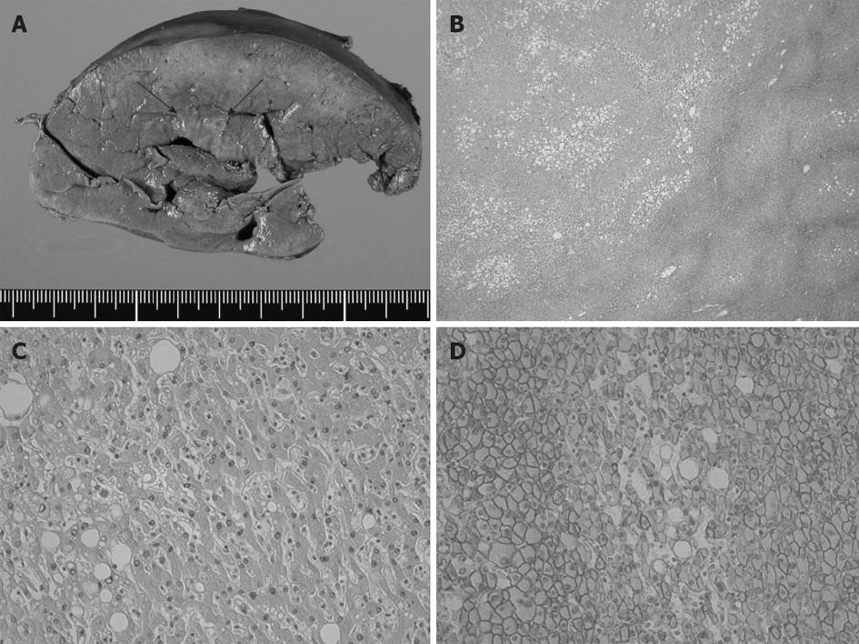
Figure 3 Pathological findings of the liver tumor.
A: Cut surface of formalin-fixed liver specimen. The tumor was unencapsulated and its border was ill-defined (arrow); B, C: Microscopically, the tumor consisted of low-grade atypical hepatocytes, without cellular mitosis or changes in cellular density and structures. Fatty deposition in the tumor cells was ubiquitously remarkable. Neither biliary structures nor portal triads were present within the tumor (hematoxylin-eosin stain, original magnification ×40 in B and ×200 in C); D: β-catenin was immunohistochemically detected on the cytomembrane. Neither aberrant nuclear nor cytoplasmic accumulations were found (original magnification ×100).
- Citation: Inaba K, Sakaguchi T, Kurachi K, Mori H, Tao H, Nakamura T, Takehara Y, Baba S, Maekawa M, Sugimura H, Konno H. Hepatocellular adenoma associated with familial adenomatous polyposis coli. World J Hepatol 2012; 4(11): 322-326
- URL: https://www.wjgnet.com/1948-5182/full/v4/i11/322.htm
- DOI: https://dx.doi.org/10.4254/wjh.v4.i11.322


