Copyright
©The Author(s) 2022.
World J Hepatol. May 27, 2022; 14(5): 1038-1046
Published online May 27, 2022. doi: 10.4254/wjh.v14.i5.1038
Published online May 27, 2022. doi: 10.4254/wjh.v14.i5.1038
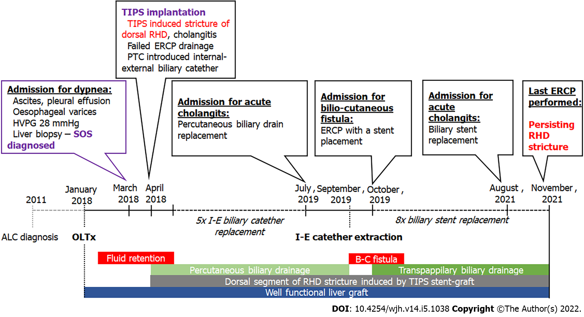
Figure 1 Timeline of the reported case.
ALC: Alcoholic liver cirrhosis; B-C: Bilio-cutaneous; I-E: Internal-external; OLTx: Orthotopic liver transplantation; RHD: Right hepatic duct; SOS: Sinusoidal obstruction syndrome.
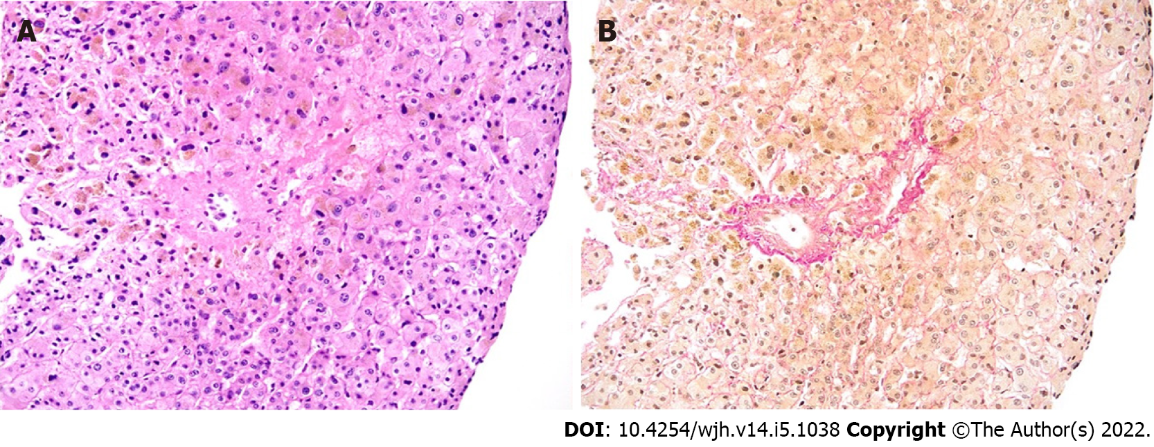
Figure 2 Histological findings in liver biopsy.
Centro lobular vein with wall edema and narrowing of the lumen by connective tissue, focal obstructive fibrosis of the surrounding sinuses; hematoxylin-eosin (A) and Elastica van Gieson (B) staining, original magnification x 100.
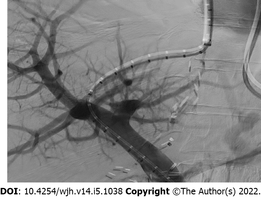
Figure 3 Transjugular intrahepatic portosystemic shunt implantation.
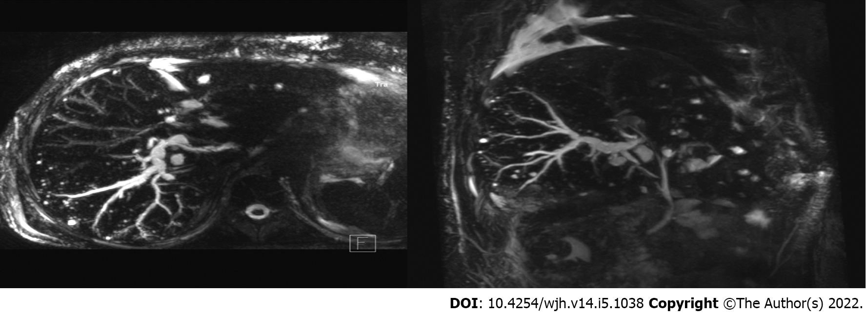
Figure 4 Magnetic resonance imaging scan after transjugular intrahepatic portosystemic shunt placement.
Dilation of dorsal branch of right hepatic duct with multiple small abscesses of the right lobe.
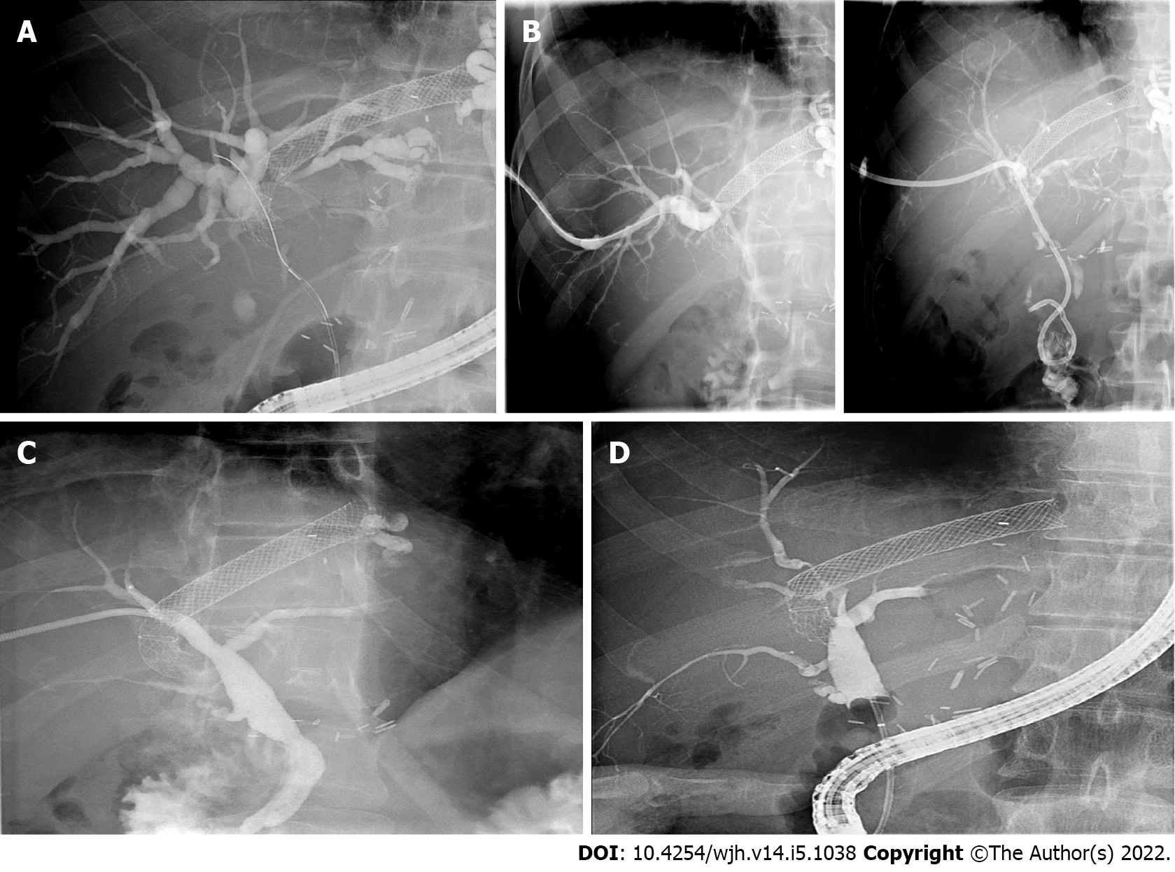
Figure 5 Images of cholangiogram.
A: Endoscopic cholangiogram. Tight stenosis in the dorsal segment of the right hepatic duct caused by the transjugular intrahepatic portosystemic shunt stent graft; stent not placed; B: Percutaneous cholangiogram. Stricture passed with a wire; external-internal catheter placed in duodenum; C: Percutaneous cholangiogram. Apparent regression of the visualized stricture with drain removed; D: Eight endoscopic cholangiograms. Persistent stricture of the right hepatic duct.
- Citation: Macinga P, Gogova D, Raupach J, Jarosova J, Janousek L, Honsova E, Taimr P, Spicak J, Novotny J, Peregrin J, Hucl T. Biliary obstruction following transjugular intrahepatic portosystemic shunt placement in a patient after liver transplantation: A case report. World J Hepatol 2022; 14(5): 1038-1046
- URL: https://www.wjgnet.com/1948-5182/full/v14/i5/1038.htm
- DOI: https://dx.doi.org/10.4254/wjh.v14.i5.1038


